


Microscope Parts & Functions
Microscope Components


What is a Microscope?
A microscope is a scientific instrument used to magnify and observe objects that are too small to be seen with the naked eye. It works by focusing light or electrons to create an enlarged image of the specimen.
Check out some of our favorite microscopes
What is a Specimen?
A specimen is a sample or example used for scientific study. It can be anything from biological tissues to materials, examined under a microscope or other instruments for analysis.
Learn More About Microscopes
What is a Compound Microscope?
A Compound Microscope is a type of optical microscope that uses multiple lenses to magnify small objects. It consists of two sets of lenses: the objective lens, which is closer to the specimen and provides the initial magnification, and the eyepiece lens, which further magnifies the image for the viewer's eye. Light passes through the specimen and is magnified by the objective lens, then further magnified by the eyepiece lens, resulting in a highly magnified image visible to the observer. Compound microscopes are commonly used in biology, medicine, and other scientific fields for viewing cells, tissues, and other small structures.
What is a Stereo Microscope?
A stereo microscope, also known as a stereoscopic or dissecting microscope, provides three-dimensional viewing of larger, opaque specimens through dual optical paths with objective lenses. It offers lower magnification (typically 5x to 40x) than compound microscopes but enhances depth perception. Ideal for tasks in biology, geology, and manufacturing, it allows comfortable, extended viewing with ergonomic adjustments.
What is the difference between Compound and Stereo Microscope?
Compound microscopes are suited for detailed examination of microscopic structures, while stereo microscopes are more appropriate for observing larger objects in three dimensions and for tasks that involve manipulation and dissection.
Stereo Microscope:
Optical System:
Uses two separate optical paths with two objective lenses to provide a stereoscopic (3D) view of larger, opaque specimens.
Provides lower magnification (typically 10x to 40x) compared to compound microscopes.
Magnification:
Offers lower magnification levels suitable for viewing objects that are larger and more three-dimensional.
Magnification is achieved through the combination of the objective lenses and sometimes adjustable zoom lenses.
Applications:
Ideal for examining larger specimens such as insects, plants, rocks, circuit boards, and other opaque objects.
Used in fields like biology, geology, entomology, electronics assembly, and manufacturing for tasks requiring manipulation and examination of objects in three dimensions.
Viewing:
Provides a stereoscopic (3D) view of the specimen, allowing for depth perception and spatial orientation.
Useful for tasks that require hand-eye coordination and manipulation of specimens.
Compound Microscope:
Optical System:
Uses multiple lenses in series (objective and eyepiece) to magnify the image of a thin, transparent specimen.
Provides high magnification (up to 1000x or more) and high resolution for viewing fine details of cells, tissues, and microorganisms.
Typically employs transmitted light illumination, where light passes through the specimen from below.
Magnification:
Capable of high magnification, which is achieved through the combination of the objective lens (typically 4x, 10x, 40x, and 100x) and the eyepiece (usually 10x).
Applications:
Ideal for observing microscopic structures, such as cells, bacteria, and tissue sections.
Commonly used in biological research, medical diagnostics, and educational settings for detailed examination of specimens.
Viewing:
Provides a flat, two-dimensional image of the specimen.
Does not provide a true stereoscopic (3D) view.
What is the difference between a Monocular, Binocular and Trinocular heads?
The terms monocular, binocular, and trinocular refer to the different types of microscope heads, each offering a distinct way of viewing the specimen.
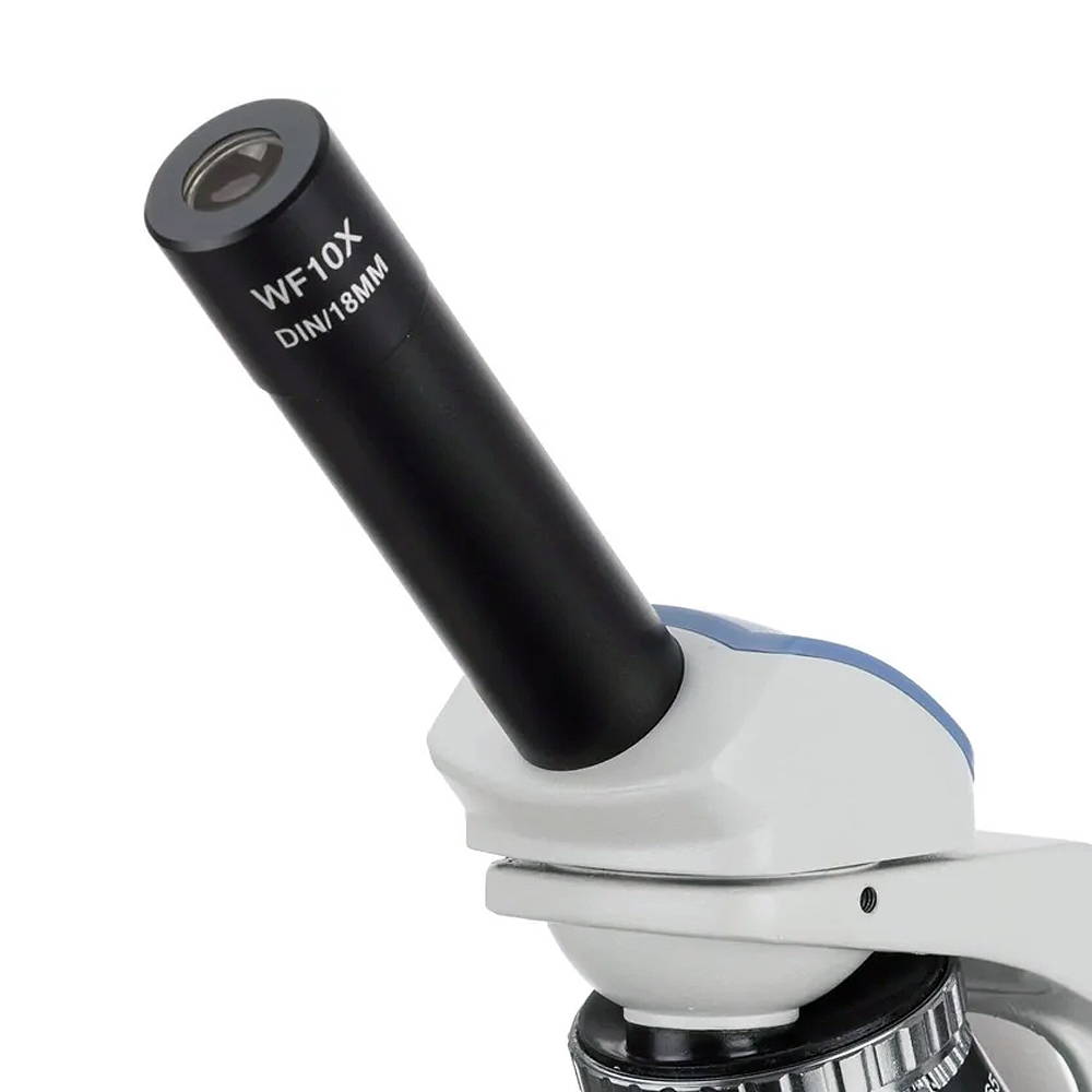
Monocular
Microscope Head
A monocular microscope head is a basic type of microscope head with a single eyepiece, ideal for cost-effective and straightforward applications. It is particularly useful in educational settings and for beginners, but it can lead to eye strain over long periods and lacks the depth perception provided by more advanced binocular and trinocular heads.
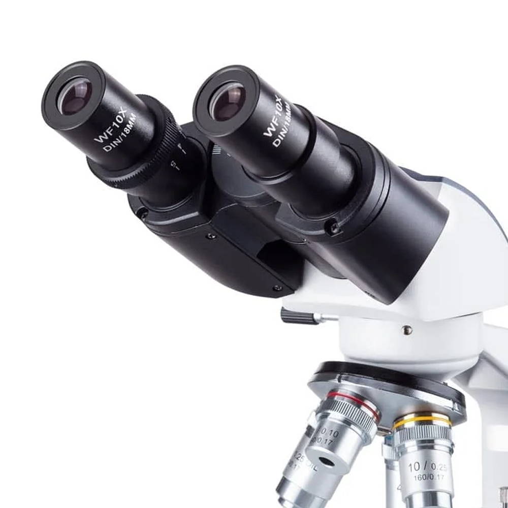
Binocular
Microscope Head
A binocular microscope head utilizes two eyepieces for simultaneous viewing with both eyes, providing enhanced comfort, depth perception, and superior image quality. Ideal for professional and research settings requiring detailed observation, its design minimizes eye strain and enhances ergonomic support compared to monocular microscopes.
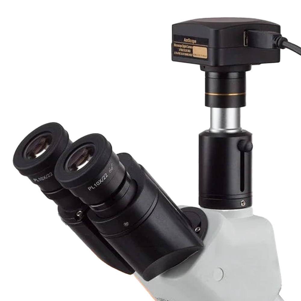
Trinocular
Microscope Head
A trinocular microscope head combines the benefits of binocular viewing with the capability to capture digital images or videos of specimens. It is particularly suited for advanced research, educational purposes, and industrial applications where precise imaging and documentation are essential.
What are Microscope Objectives?
Microscope objectives are vital lenses that determine the magnification, resolution, and quality of the images produced by a microscope. They come in various types and magnifications, each suited for different applications and levels of detail, making them indispensable in scientific research, medical diagnostics, and educational settings.
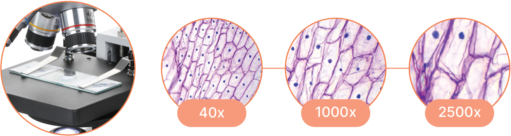
How do you calculate Magnification?
Compound Magnification is calculated by multiplying the magnification of the objective lens by the magnification of the eyepiece.
Total Magnification = Objective Magnification X Eyepiece Magnification
What is Magnification?
Magnification is the process of enlarging the appearance of an object, making it look bigger than its actual size. In optics, it is the ratio of the size of the image produced by a lens or microscope to the actual size of the object being viewed.
How Does it Work?
Magnification works by bending light through lenses or using digital technology to enlarge the appearance of an object, allowing for detailed observation and analysis.
What is a Darkfield or Phase Contrast Microscope?
Darkfield Microscope
A darkfield microscope is a type of optical microscope that provides high contrast images of unstained specimens by using scattered light. The specimen appears bright against a dark background
How It Works
Light Source: Illuminates the specimen.
Condenser: Directs light in a hollow cone so it doesn’t enter the objective lens directly.
Specimen Interaction: Light scatters upon interacting with the specimen.
Objective Lens: Collects the scattered light.
Image Formation: The scattered light forms a bright image of the specimen on a dark background.
Uses
Live Cell Imaging: Observing live, unstained cells.
Particle Detection: Detecting small particles and material defects.
Forensic Science: Examining fibers and hairs.
Clinical Diagnosis: Detecting bacteria like spirochetes in blood samples.
Advantages
High Contrast: Ideal for transparent and unstained specimens.
Non-destructive: No staining required.
Limitations
Qualitative: Mostly for qualitative analysis, not detailed structure.
Specimen Thickness: Best for thin specimens.
Maintenance: Requires precise alignment and maintenance.
Phase Contrast Microscope
A phase contrast microscope is an optical microscope designed to enhance the contrast of transparent and colorless specimens without the need for staining. It works by exploiting differences in the refractive index of different parts of the specimen, transforming these differences into variations in light intensity.
How It Works
Light Source: Provides illumination.
Annular Diaphragm: Creates a hollow cone of light.
Specimen Interaction: Light undergoes phase shifts passing through the specimen.
Phase Plate: Alters the phase of direct light differently from scattered light.
Image Formation: Interference between direct and scattered light creates a high-contrast image.
Uses
Cell Biology: Observing live cells and structures.
Microbiology: Studying microorganisms.
Medical Diagnosis: Examining blood cells and tissues.
Research: Investigating tissue slices and cellular components.
Advantages
Enhanced Contrast: For transparent specimens.
Non-destructive: No staining needed.
Detailed Observations: Reveals internal structures.
Limitations
Halo Effect: May produce halos around specimens.
Complex Setup: Requires specific optics.
Light Loss: Reduces light intensity.
Precision Optics
Witness the microscopic world in stunning detail with our high-quality optics. Every slide comes to life with crystal-clear clarity, allowing you to delve into the intricacies of biology, chemistry, and beyond.
Innovative Illumination
Illuminate your subjects with brilliance. Our microscopes feature advanced lighting technologies, providing the perfect balance for optimal observation, even in low-light conditions.
User-Friendly Design
Navigate effortlessly through magnification levels and focus adjustments. Our microscopes feature intuitive controls, allowing you to concentrate on your research without the hassle of complicated settings.