


AmScope T520B Trinocular Biological Microscope with 2000X Magnification and 20W Incandescent Light Source 3D Mechanical Stage AmScope T520B-M Digital Enabled Trinocular Biological Microscope C-Mount with 2000X Magnification and 20W Incandescent Light Source includes a USB 2.0 1.3MP Camera 3D Mechanical Stage AmScope T520B-3M Digital Enabled Trinocular Biological Microscope C-Mount with 2000X Magnification and 20W Incandescent Light Source includes a USB 2.0 3MP Camera 3D Mechanical Stage AmScope T520B-5M Digital Enabled Trinocular Biological Microscope C-Mount with 2000X Magnification and 20W Incandescent Light Source includes a USB 2.0 5MP Camera 3D Mechanical Stage AmScope T520B-8M Digital Enabled Trinocular Biological Microscope C-Mount with 2000X Magnification and 20W Incandescent Light Source includes a USB 2.0 8MP Camera 3D Mechanical Stage AmScope T520B-9M Digital Enabled Trinocular Biological Microscope C-Mount with 2000X Magnification and 20W Incandescent Light Source includes a USB 2.0 9MP Camera 3D Mechanical Stage AmScope T520B-10M Digital Enabled Trinocular Biological Microscope C-Mount with 2000X Magnification and 20W Incandescent Light Source includes a USB 2.0 10MP Camera 3D Mechanical Stage AmScope T520B-3M3 Digital Enabled Trinocular Biological Microscope C-Mount with 2000X Magnification and 20W Incandescent Light Source includes a USB 3.0 3MP Camera 3D Mechanical Stage AmScope T520B-5M3 Digital Enabled Trinocular Biological Microscope C-Mount with 2000X Magnification and 20W Incandescent Light Source includes a USB 3.0 5MP Camera 3D Mechanical Stage AmScope T520B-10M3 Digital Enabled Trinocular Biological Microscope C-Mount with 2000X Magnification and 20W Incandescent Light Source includes a USB 3.0 10MP Camera 3D Mechanical Stage AmScope T520B-14M3 Digital Enabled Trinocular Biological Microscope C-Mount with 2000X Magnification and 20W Incandescent Light Source includes a USB 3.0 14MP Camera 3D Mechanical Stage AmScope T520B-18M3 Digital Enabled Trinocular Biological Microscope C-Mount with 2000X Magnification and 20W Incandescent Light Source includes a USB 3.0 18MP Camera 3D Mechanical Stage AmScope T520 Trinocular Biological Microscope with 1000X Magnification and 20W Incandescent Light Source 3D Mechanical Stage AmScope T520-M Digital Enabled Trinocular Biological Microscope C-Mount with 1000X Magnification and 20W Incandescent Light Source includes a USB 2.0 1.3MP Camera 3D Mechanical Stage AmScope T520-3M Digital Enabled Trinocular Biological Microscope C-Mount with 1000X Magnification and 20W Incandescent Light Source includes a USB 2.0 3MP Camera 3D Mechanical Stage AmScope T520-5M Digital Enabled Trinocular Biological Microscope C-Mount with 1000X Magnification and 20W Incandescent Light Source includes a USB 2.0 5MP Camera 3D Mechanical Stage AmScope T520-8M Digital Enabled Trinocular Biological Microscope C-Mount with 1000X Magnification and 20W Incandescent Light Source includes a USB 2.0 8MP Camera 3D Mechanical Stage AmScope T520-9M Digital Enabled Trinocular Biological Microscope C-Mount with 1000X Magnification and 20W Incandescent Light Source includes a USB 2.0 9MP Camera 3D Mechanical Stage AmScope T520-10M Digital Enabled Trinocular Biological Microscope C-Mount with 1000X Magnification and 20W Incandescent Light Source includes a USB 2.0 10MP Camera 3D Mechanical Stage AmScope T520-3M3 Digital Enabled Trinocular Biological Microscope C-Mount with 1000X Magnification and 20W Incandescent Light Source includes a USB 3.0 3MP Camera 3D Mechanical Stage AmScope T520-5M3 Digital Enabled Trinocular Biological Microscope C-Mount with 1000X Magnification and 20W Incandescent Light Source includes a USB 3.0 5MP Camera 3D Mechanical Stage AmScope T520-10M3 Digital Enabled Trinocular Biological Microscope C-Mount with 1000X Magnification and 20W Incandescent Light Source includes a USB 3.0 10MP Camera 3D Mechanical Stage AmScope T520-14M3 Digital Enabled Trinocular Biological Microscope C-Mount with 1000X Magnification and 20W Incandescent Light Source includes a USB 3.0 14MP Camera 3D Mechanical Stage AmScope T520-18M3 Digital Enabled Trinocular Biological Microscope C-Mount with 1000X Magnification and 20W Incandescent Light Source includes a USB 3.0 18MP Camera 3D Mechanical Stage AmScope T520A Trinocular Biological Microscope with 1600X Magnification and 20W Incandescent Light Source 3D Mechanical Stage AmScope T520A-M Digital Enabled Trinocular Biological Microscope C-Mount with 1600X Magnification and 20W Incandescent Light Source includes a USB 2.0 1.3MP Camera 3D Mechanical Stage AmScope T520A-3M Digital Enabled Trinocular Biological Microscope C-Mount with 1600X Magnification and 20W Incandescent Light Source includes a USB 2.0 3MP Camera 3D Mechanical Stage AmScope T520A-5M Digital Enabled Trinocular Biological Microscope C-Mount with 1600X Magnification and 20W Incandescent Light Source includes a USB 2.0 5MP Camera 3D Mechanical Stage AmScope T520A-8M Digital Enabled Trinocular Biological Microscope C-Mount with 1600X Magnification and 20W Incandescent Light Source includes a USB 2.0 8MP Camera 3D Mechanical Stage AmScope T520A-9M Digital Enabled Trinocular Biological Microscope C-Mount with 1600X Magnification and 20W Incandescent Light Source includes a USB 2.0 9MP Camera 3D Mechanical Stage AmScope T520A-10M Digital Enabled Trinocular Biological Microscope C-Mount with 1600X Magnification and 20W Incandescent Light Source includes a USB 2.0 10MP Camera 3D Mechanical Stage AmScope T520A-3M3 Digital Enabled Trinocular Biological Microscope C-Mount with 1600X Magnification and 20W Incandescent Light Source includes a USB 3.0 3MP Camera 3D Mechanical Stage AmScope T520A-5M3 Digital Enabled Trinocular Biological Microscope C-Mount with 1600X Magnification and 20W Incandescent Light Source includes a USB 3.0 5MP Camera 3D Mechanical Stage AmScope T520A-10M3 Digital Enabled Trinocular Biological Microscope C-Mount with 1600X Magnification and 20W Incandescent Light Source includes a USB 3.0 10MP Camera 3D Mechanical Stage AmScope T520A-14M3 Digital Enabled Trinocular Biological Microscope C-Mount with 1600X Magnification and 20W Incandescent Light Source includes a USB 3.0 14MP Camera 3D Mechanical Stage AmScope T520A-18M3 Digital Enabled Trinocular Biological Microscope C-Mount with 1600X Magnification and 20W Incandescent Light Source includes a USB 3.0 18MP Camera 3D Mechanical Stage AmScope T520C Trinocular Biological Microscope with 2500X Magnification and 20W Incandescent Light Source 3D Mechanical Stage AmScope T520C-M Digital Enabled Trinocular Biological Microscope C-Mount with 2500X Magnification and 20W Incandescent Light Source includes a USB 2.0 1.3MP Camera 3D Mechanical Stage AmScope T520C-3M Digital Enabled Trinocular Biological Microscope C-Mount with 2500X Magnification and 20W Incandescent Light Source includes a USB 2.0 3MP Camera 3D Mechanical Stage AmScope T520C-5M Digital Enabled Trinocular Biological Microscope C-Mount with 2500X Magnification and 20W Incandescent Light Source includes a USB 2.0 5MP Camera 3D Mechanical Stage AmScope T520C-8M Digital Enabled Trinocular Biological Microscope C-Mount with 2500X Magnification and 20W Incandescent Light Source includes a USB 2.0 8MP Camera 3D Mechanical Stage AmScope T520C-9M Digital Enabled Trinocular Biological Microscope C-Mount with 2500X Magnification and 20W Incandescent Light Source includes a USB 2.0 9MP Camera 3D Mechanical Stage AmScope T520C-10M Digital Enabled Trinocular Biological Microscope C-Mount with 2500X Magnification and 20W Incandescent Light Source includes a USB 2.0 10MP Camera 3D Mechanical Stage AmScope T520C-3M3 Digital Enabled Trinocular Biological Microscope C-Mount with 2500X Magnification and 20W Incandescent Light Source includes a USB 3.0 3MP Camera 3D Mechanical Stage AmScope T520C-5M3 Digital Enabled Trinocular Biological Microscope C-Mount with 2500X Magnification and 20W Incandescent Light Source includes a USB 3.0 5MP Camera 3D Mechanical Stage AmScope T520C-10M3 Digital Enabled Trinocular Biological Microscope C-Mount with 2500X Magnification and 20W Incandescent Light Source includes a USB 3.0 10MP Camera 3D Mechanical Stage AmScope T520C-14M3 Digital Enabled Trinocular Biological Microscope C-Mount with 2500X Magnification and 20W Incandescent Light Source includes a USB 3.0 14MP Camera 3D Mechanical Stage AmScope T520C-18M3 Digital Enabled Trinocular Biological Microscope C-Mount with 2500X Magnification and 20W Incandescent Light Source includes a USB 3.0 18MP Camera 3D Mechanical Stage
Key Features
- The T520 provides high quality optics and precision mechanics for use in classrooms and clinics.
- Professional features include a Siedentopf trinocular head for precise adjustments, a 2-layer mechanical stage with low-position controls, and coaxial coarse and fine focus for ergonomics and an efficient workflow.
- Halogen lighting reduces eye-strain for prolonged use.
- The adjustable trinocular photo port allows you to easily mount a camera for real-time monitoring or capturing photos and videos.
- Monitor and capture photos and videos with the included 1.3MP USB 2.0 camera. Professional microscopy software for Windows, Mac, and Linux provide tools for image processing, measuring, and more.
- The T520 provides high quality optics and precision mechanics for use in classrooms and clinics.
- Professional features include a Siedentopf trinocular head for precise adjustments, a 2-layer mechanical stage with low-position controls, and coaxial coarse and fine focus for ergonomics and an efficient workflow.
- Halogen lighting reduces eye-strain for prolonged use.
- The adjustable trinocular photo port allows you to easily mount a camera for real-time monitoring or capturing photos and videos.
- Monitor and capture photos and videos with the included 3MP USB 2.0 camera. Professional microscopy software for Windows, Mac, and Linux provide tools for image processing, measuring, and more.
- The T520 provides high quality optics and precision mechanics for use in classrooms and clinics.
- Professional features include a Siedentopf trinocular head for precise adjustments, a 2-layer mechanical stage with low-position controls, and coaxial coarse and fine focus for ergonomics and an efficient workflow.
- Halogen lighting reduces eye-strain for prolonged use.
- The adjustable trinocular photo port allows you to easily mount a camera for real-time monitoring or capturing photos and videos.
- Monitor and capture photos and videos with the included 5MP USB 2.0 camera. Professional microscopy software for Windows, Mac, and Linux provide tools for image processing, measuring, and more.
- The T520 provides high quality optics and precision mechanics for use in classrooms and clinics.
- Professional features include a Siedentopf trinocular head for precise adjustments, a 2-layer mechanical stage with low-position controls, and coaxial coarse and fine focus for ergonomics and an efficient workflow.
- Halogen lighting reduces eye-strain for prolonged use.
- The adjustable trinocular photo port allows you to easily mount a camera for real-time monitoring or capturing photos and videos.
- Monitor and capture photos and videos with the included 8MP USB 2.0 camera. Professional microscopy software for Windows and Mac provide tools for image processing, measuring, and more.
- The T520 provides high quality optics and precision mechanics for use in classrooms and clinics.
- Professional features include a Siedentopf trinocular head for precise adjustments, a 2-layer mechanical stage with low-position controls, and coaxial coarse and fine focus for ergonomics and an efficient workflow.
- Halogen lighting reduces eye-strain for prolonged use.
- The adjustable trinocular photo port allows you to easily mount a camera for real-time monitoring or capturing photos and videos.
- Monitor and capture photos and videos with the included 9MP USB 2.0 camera. Professional microscopy software for Windows, Mac, and Linux provide tools for image processing, measuring, and more.
- The T520 provides high quality optics and precision mechanics for use in classrooms and clinics.
- Professional features include a Siedentopf trinocular head for precise adjustments, a 2-layer mechanical stage with low-position controls, and coaxial coarse and fine focus for ergonomics and an efficient workflow.
- Halogen lighting reduces eye-strain for prolonged use.
- The adjustable trinocular photo port allows you to easily mount a camera for real-time monitoring or capturing photos and videos.
- Monitor and capture photos and videos with the included 10MP USB 2.0 camera. Professional microscopy software for Windows, Mac, and Linux provide tools for image processing, measuring, and more.
- The T520 provides high quality optics and precision mechanics for use in classrooms and clinics.
- Professional features include a Siedentopf trinocular head for precise adjustments, a 2-layer mechanical stage with low-position controls, and coaxial coarse and fine focus for ergonomics and an efficient workflow.
- Halogen lighting reduces eye-strain for prolonged use.
- The adjustable trinocular photo port allows you to easily mount a camera for real-time monitoring or capturing photos and videos.
- Monitor and capture photos and videos with the included 3MP USB 3.0 camera. Professional microscopy software for Windows, Mac, and Linux provide tools for image processing, measuring, and more.
- The T520 provides high quality optics and precision mechanics for use in classrooms and clinics.
- Professional features include a Siedentopf trinocular head for precise adjustments, a 2-layer mechanical stage with low-position controls, and coaxial coarse and fine focus for ergonomics and an efficient workflow.
- Halogen lighting reduces eye-strain for prolonged use.
- The adjustable trinocular photo port allows you to easily mount a camera for real-time monitoring or capturing photos and videos.
- Monitor and capture photos and videos with the included 5MP USB 3.0 camera. Professional microscopy software for Windows, Mac, and Linux provide tools for image processing, measuring, and more.
- The T520 provides high quality optics and precision mechanics for use in classrooms and clinics.
- Professional features include a Siedentopf trinocular head for precise adjustments, a 2-layer mechanical stage with low-position controls, and coaxial coarse and fine focus for ergonomics and an efficient workflow.
- Halogen lighting reduces eye-strain for prolonged use.
- The adjustable trinocular photo port allows you to easily mount a camera for real-time monitoring or capturing photos and videos.
- Monitor and capture photos and videos with the included 10MP USB 3.0 camera. Professional microscopy software for Windows, Mac, and Linux provide tools for image processing, measuring, and more.
- The T520 provides high quality optics and precision mechanics for use in classrooms and clinics.
- Professional features include a Siedentopf trinocular head for precise adjustments, a 2-layer mechanical stage with low-position controls, and coaxial coarse and fine focus for ergonomics and an efficient workflow.
- Halogen lighting reduces eye-strain for prolonged use.
- The adjustable trinocular photo port allows you to easily mount a camera for real-time monitoring or capturing photos and videos.
- Monitor and capture photos and videos with the included 14MP USB 3.0 camera. Professional microscopy software for Windows, Mac, and Linux provide tools for image processing, measuring, and more.
- Free Upgrade: The 18MP camera has been replaced by our new 20MP camera!
- The T520 provides high quality optics and precision mechanics for use in classrooms and clinics.
- Professional features include a Siedentopf trinocular head for precise adjustments, a 2-layer mechanical stage with low-position controls, and coaxial coarse and fine focus for ergonomics and an efficient workflow.
- Halogen lighting reduces eye-strain for prolonged use.
- The adjustable trinocular photo port allows you to easily mount a camera for real-time monitoring or capturing photos and videos.
- Monitor and capture photos and videos with the included 18MP 20MP USB 3.0 camera. Professional microscopy software for Windows, Mac, and Linux provide tools for image processing, measuring, and more.
- The T520 provides high quality optics and precision mechanics for use in classrooms and clinics.
- Professional features include a Siedentopf trinocular head for precise adjustments, a 2-layer mechanical stage with low-position controls, and coaxial coarse and fine focus for ergonomics and an efficient workflow.
- Halogen lighting reduces eye-strain for prolonged use.
- The adjustable trinocular photo port allows you to easily mount a camera for real-time monitoring or capturing photos and videos.
- Monitor and capture photos and videos with the included 1.3MP USB 2.0 camera. Professional microscopy software for Windows, Mac, and Linux provide tools for image processing, measuring, and more.
- The T520 provides high quality optics and precision mechanics for use in classrooms and clinics.
- Professional features include a Siedentopf trinocular head for precise adjustments, a 2-layer mechanical stage with low-position controls, and coaxial coarse and fine focus for ergonomics and an efficient workflow.
- Halogen lighting reduces eye-strain for prolonged use.
- The adjustable trinocular photo port allows you to easily mount a camera for real-time monitoring or capturing photos and videos.
- Monitor and capture photos and videos with the included 3MP USB 2.0 camera. Professional microscopy software for Windows, Mac, and Linux provide tools for image processing, measuring, and more.
- The T520 provides high quality optics and precision mechanics for use in classrooms and clinics.
- Professional features include a Siedentopf trinocular head for precise adjustments, a 2-layer mechanical stage with low-position controls, and coaxial coarse and fine focus for ergonomics and an efficient workflow.
- Halogen lighting reduces eye-strain for prolonged use.
- The adjustable trinocular photo port allows you to easily mount a camera for real-time monitoring or capturing photos and videos.
- Monitor and capture photos and videos with the included 5MP USB 2.0 camera. Professional microscopy software for Windows, Mac, and Linux provide tools for image processing, measuring, and more.
- The T520 provides high quality optics and precision mechanics for use in classrooms and clinics.
- Professional features include a Siedentopf trinocular head for precise adjustments, a 2-layer mechanical stage with low-position controls, and coaxial coarse and fine focus for ergonomics and an efficient workflow.
- Halogen lighting reduces eye-strain for prolonged use.
- The adjustable trinocular photo port allows you to easily mount a camera for real-time monitoring or capturing photos and videos.
- Monitor and capture photos and videos with the included 8MP USB 2.0 camera. Professional microscopy software for Windows and Mac provide tools for image processing, measuring, and more.
- The T520 provides high quality optics and precision mechanics for use in classrooms and clinics.
- Professional features include a Siedentopf trinocular head for precise adjustments, a 2-layer mechanical stage with low-position controls, and coaxial coarse and fine focus for ergonomics and an efficient workflow.
- Halogen lighting reduces eye-strain for prolonged use.
- The adjustable trinocular photo port allows you to easily mount a camera for real-time monitoring or capturing photos and videos.
- Monitor and capture photos and videos with the included 9MP USB 2.0 camera. Professional microscopy software for Windows, Mac, and Linux provide tools for image processing, measuring, and more.
- The T520 provides high quality optics and precision mechanics for use in classrooms and clinics.
- Professional features include a Siedentopf trinocular head for precise adjustments, a 2-layer mechanical stage with low-position controls, and coaxial coarse and fine focus for ergonomics and an efficient workflow.
- Halogen lighting reduces eye-strain for prolonged use.
- The adjustable trinocular photo port allows you to easily mount a camera for real-time monitoring or capturing photos and videos.
- Monitor and capture photos and videos with the included 10MP USB 2.0 camera. Professional microscopy software for Windows, Mac, and Linux provide tools for image processing, measuring, and more.
- The T520 provides high quality optics and precision mechanics for use in classrooms and clinics.
- Professional features include a Siedentopf trinocular head for precise adjustments, a 2-layer mechanical stage with low-position controls, and coaxial coarse and fine focus for ergonomics and an efficient workflow.
- Halogen lighting reduces eye-strain for prolonged use.
- The adjustable trinocular photo port allows you to easily mount a camera for real-time monitoring or capturing photos and videos.
- Monitor and capture photos and videos with the included 3MP USB 3.0 camera. Professional microscopy software for Windows, Mac, and Linux provide tools for image processing, measuring, and more.
- The T520 provides high quality optics and precision mechanics for use in classrooms and clinics.
- Professional features include a Siedentopf trinocular head for precise adjustments, a 2-layer mechanical stage with low-position controls, and coaxial coarse and fine focus for ergonomics and an efficient workflow.
- Halogen lighting reduces eye-strain for prolonged use.
- The adjustable trinocular photo port allows you to easily mount a camera for real-time monitoring or capturing photos and videos.
- Monitor and capture photos and videos with the included 5MP USB 3.0 camera. Professional microscopy software for Windows, Mac, and Linux provide tools for image processing, measuring, and more.
- The T520 provides high quality optics and precision mechanics for use in classrooms and clinics.
- Professional features include a Siedentopf trinocular head for precise adjustments, a 2-layer mechanical stage with low-position controls, and coaxial coarse and fine focus for ergonomics and an efficient workflow.
- Halogen lighting reduces eye-strain for prolonged use.
- The adjustable trinocular photo port allows you to easily mount a camera for real-time monitoring or capturing photos and videos.
- Monitor and capture photos and videos with the included 10MP USB 3.0 camera. Professional microscopy software for Windows, Mac, and Linux provide tools for image processing, measuring, and more.
- The T520 provides high quality optics and precision mechanics for use in classrooms and clinics.
- Professional features include a Siedentopf trinocular head for precise adjustments, a 2-layer mechanical stage with low-position controls, and coaxial coarse and fine focus for ergonomics and an efficient workflow.
- Halogen lighting reduces eye-strain for prolonged use.
- The adjustable trinocular photo port allows you to easily mount a camera for real-time monitoring or capturing photos and videos.
- Monitor and capture photos and videos with the included 14MP USB 3.0 camera. Professional microscopy software for Windows, Mac, and Linux provide tools for image processing, measuring, and more.
- Free Upgrade: The 18MP camera has been replaced by our new 20MP camera!
- The T520 provides high quality optics and precision mechanics for use in classrooms and clinics.
- Professional features include a Siedentopf trinocular head for precise adjustments, a 2-layer mechanical stage with low-position controls, and coaxial coarse and fine focus for ergonomics and an efficient workflow.
- Halogen lighting reduces eye-strain for prolonged use.
- The adjustable trinocular photo port allows you to easily mount a camera for real-time monitoring or capturing photos and videos.
- Monitor and capture photos and videos with the included 18MP 20MP USB 3.0 camera. Professional microscopy software for Windows, Mac, and Linux provide tools for image processing, measuring, and more.
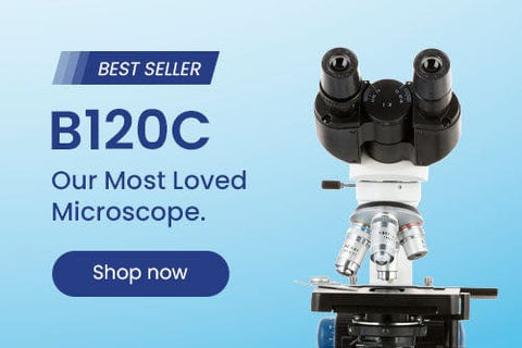
The T520 is a medium-sized compound microscope designed for biological applications. Ideally suited for classrooms and veterinary practices, this model provides essential functionality with a solid build.
The trinocular head accommodates viewing through eyepieces and through the top-mounted photo port. The binocular viewing tubes use Siedentopf adjustment to easily fit any user. A diopter is available on the left ocular tube to compensate for asymmetry, and the ocular tubes are angled at 30° to comfortably accommodate a seated position without neck strain. The head has a top-mounted, height-adjustable 23mm photo port for mounting optional cameras. C-mount cameras can be paired with various reduction lenses for mounting to the port and optimizing field-of-view.
The objective turret provides instant access to 4 magnification levels to easily focus in on minute details from 40X to 1000X. Additional 20X eyepieces expand the maximum magnification to 2000X with 8 unique levels. This covers magnifications needed for studying hair follicles, cells, and bacteria. The turret's reversed orientation provides more working room above the stage. These high-quality lenses are achromatically corrected to improve resolution and color accuracy.
The 2-layer mechanical stage provides smooth and precise movement for examination of specimen slides. The stage's low-position controls are conveniently placed near the coaxial focus knobs for a streamlined workflow. Examining specimen slides is intuitive and precise for users at any skill level.
The halogen sub-stage lighting provides bright illumination with excellent color rendering. The warm lighting is better suited for prolonged use, causing less eyestrain than LED and fluorescent lighting. The Abbe light condenser has a high 1.25 numerical aperture for oil-immersion, and an iris diaphragm to optimize contrast and depth-of-field. The condenser holder uses rack-and-pinion movement for precise height adjustment, and has centering screws for properly aligning the condenser.
Specifications
| Optical System | Finite-conjugate |
| Mechanical Tube Length | 160mm |
| Head | Trinocular, 30° incline, 360° rotatable |
| Interpupillary Adjustment | Siedentopf compensation-free gemel, 55-75mm |
| Ocular Diameter | 23mm |
| Eyepieces | 10X, 20X |
| Photo Port | Height-adjustable 23mm tube |
| Objective Lenses | DIN standard |
| Objective Parfocal Distance | 45mm |
| Objective Mounting Thread | RMS 20.32mm |
| Objective Turret | reversed, quadruple |
| Focusing System | Coaxial coarse and fine focus, tension control |
| Division of Fine Focus | 0.002mm |
| Stage Design | Double-layer with caliper |
| Stage Dimensions | 140mm x 132mm |
| X-Y Translation Range | 75mm x 30mm |
| Transmitted Illumination | 20W halogen |
| Condenser | Abbe condenser with iris diaphragm, NA1.25 |
| Sub-stage Condenser-holder | rack and pinion |
| Power | 85-265VAC 50/60Hz wide-band |
Objective Lenses
| Magnification | Corrections | NA | Immersion Medium | Cover-glass Thickness |
|---|---|---|---|---|
| 4X | achromatic | 0.10 | — | 0.17mm |
| 10X | achromatic | 0.25 | — | 0.17mm |
| 40X | achromatic | 0.65 | — | 0.17mm |
| 100X | achromatic | 1.25 | oil | 0.17mm |
Packing List:
The T520 is a medium-sized compound microscope designed for biological applications. Ideally suited for classrooms and veterinary practices, this model provides essential functionality with a solid build.
The trinocular head accommodates viewing through eyepieces and through the top-mounted photo port. The binocular viewing tubes use Siedentopf adjustment to easily fit any user. A diopter is available on the left ocular tube to compensate for asymmetry, and the ocular tubes are angled at 30° to comfortably accommodate a seated position without neck strain. The head has a top-mounted, height-adjustable 23mm photo port for mounting optional cameras. C-mount cameras can be paired with various reduction lenses for mounting to the port and optimizing field-of-view.
The objective turret provides instant access to 4 magnification levels to easily focus in on minute details from 40X to 1000X. Additional 20X eyepieces expand the maximum magnification to 2000X with 8 unique levels. This covers magnifications needed for studying hair follicles, cells, and bacteria. The turret's reversed orientation provides more working room above the stage. These high-quality lenses are achromatically corrected to improve resolution and color accuracy.
The 2-layer mechanical stage provides smooth and precise movement for examination of specimen slides. The stage's low-position controls are conveniently placed near the coaxial focus knobs for a streamlined workflow. Examining specimen slides is intuitive and precise for users at any skill level.
The halogen sub-stage lighting provides bright illumination with excellent color rendering. The warm lighting is better suited for prolonged use, causing less eyestrain than LED and fluorescent lighting. The Abbe light condenser has a high 1.25 numerical aperture for oil-immersion, and an iris diaphragm to optimize contrast and depth-of-field. The condenser holder uses rack-and-pinion movement for precise height adjustment, and has centering screws for properly aligning the condenser.
Monitor and capture photos and videos with the included 1.3MP USB2.0 camera. The C-mount camera is specially designed for use with microscopes, allowing you to watch live images, and to record photos or videos on your computer. This ability to view microscopic images on your computer reduces eye-strain, and allows groups of people to view images at the same time. A 0.37X reduction lens is included for use with 23mm-30.5mm ports to capture more of the microscope's field-of-view. Our professional software for Windows provides a wide assortment of capture and photo-editing functions including color correction, time-lapse capture, image stitching, and a full complement of measuring tools. A lite version of our software is available for Mac and Linux with essential functionality for capturing photos and videos.
Specifications
| Optical System | Finite-conjugate |
| Mechanical Tube Length | 160mm |
| Head | Trinocular, 30° incline, 360° rotatable |
| Interpupillary Adjustment | Siedentopf compensation-free gemel, 55-75mm |
| Ocular Diameter | 23mm |
| Eyepieces | 10X, 20X |
| Photo Port | Height-adjustable 23mm tube |
| Objective Lenses | DIN standard |
| Objective Parfocal Distance | 45mm |
| Objective Mounting Thread | RMS 20.32mm |
| Objective Turret | reversed, quadruple |
| Focusing System | Coaxial coarse and fine focus, tension control |
| Division of Fine Focus | 0.002mm |
| Stage Design | Double-layer with caliper |
| Stage Dimensions | 140mm x 132mm |
| X-Y Translation Range | 75mm x 30mm |
| Transmitted Illumination | 20W halogen |
| Condenser | Abbe condenser with iris diaphragm, NA1.25 |
| Sub-stage Condenser-holder | rack and pinion |
| Power | 85-265VAC 50/60Hz wide-band |
Objective Lenses
| Magnification | Corrections | NA | Immersion Medium | Cover-glass Thickness |
|---|---|---|---|---|
| 4X | achromatic | 0.10 | — | 0.17mm |
| 10X | achromatic | 0.25 | — | 0.17mm |
| 40X | achromatic | 0.65 | — | 0.17mm |
| 100X | achromatic | 1.25 | oil | 0.17mm |
| Camera Specifications | |
| Sensor Type | CMOS |
| Sensor Optical Format | 1/3" |
| Active Pixels | 1.3M (1280 x 1024) |
| Pixel Size | 3.6µm x 3.6µm |
| Active Sensor Area | 4.60mm x 3.70mm |
| Shutter | electronic rolling shutter |
| Sensitivity | 1.0V/Lux-sec |
| Spectral Response | 380-650nm with IR-cut filter |
| Capture Resolution and Maximum Framerate | 15fps @ 1280x1024 26fps @ 640x512 50fps @ 320x256 |
| Connectivity | USB 2.0 |
| Lens-mount | C-mount |
| Compensating Lens | 0.37X |
| Power | 5VDC over USB |
Software
| OS Requirements | Windows (32/64 bit) XP/Vista/7/8/10, Mac OS 10.8+, Linux kernel 3.13+ | ||||||||||||||||||
|---|---|---|---|---|---|---|---|---|---|---|---|---|---|---|---|---|---|---|---|
| Hardware Requirements | Intel Core2 2.8GHz or comparable processor, 4GB RAM | ||||||||||||||||||
| Features |
|
Packing List:
The T520 is a medium-sized compound microscope designed for biological applications. Ideally suited for classrooms and veterinary practices, this model provides essential functionality with a solid build.
The trinocular head accommodates viewing through eyepieces and through the top-mounted photo port. The binocular viewing tubes use Siedentopf adjustment to easily fit any user. A diopter is available on the left ocular tube to compensate for asymmetry, and the ocular tubes are angled at 30° to comfortably accommodate a seated position without neck strain. The head has a top-mounted, height-adjustable 23mm photo port for mounting optional cameras. C-mount cameras can be paired with various reduction lenses for mounting to the port and optimizing field-of-view.
The objective turret provides instant access to 4 magnification levels to easily focus in on minute details from 40X to 1000X. Additional 20X eyepieces expand the maximum magnification to 2000X with 8 unique levels. This covers magnifications needed for studying hair follicles, cells, and bacteria. The turret's reversed orientation provides more working room above the stage. These high-quality lenses are achromatically corrected to improve resolution and color accuracy.
The 2-layer mechanical stage provides smooth and precise movement for examination of specimen slides. The stage's low-position controls are conveniently placed near the coaxial focus knobs for a streamlined workflow. Examining specimen slides is intuitive and precise for users at any skill level.
The halogen sub-stage lighting provides bright illumination with excellent color rendering. The warm lighting is better suited for prolonged use, causing less eyestrain than LED and fluorescent lighting. The Abbe light condenser has a high 1.25 numerical aperture for oil-immersion, and an iris diaphragm to optimize contrast and depth-of-field. The condenser holder uses rack-and-pinion movement for precise height adjustment, and has centering screws for properly aligning the condenser.
Monitor and capture photos and videos with the included 3.0MP USB2.0 camera. The C-mount camera is specially designed for use with microscopes, allowing you to watch live images, and to record photos or videos on your computer. This ability to view microscopic images on your computer reduces eye-strain, and allows groups of people to view images at the same time. A 0.5X reduction lens is included for use with 23mm-30.5mm ports to capture more of the microscope's field-of-view. Our professional software for Windows provides a wide assortment of capture and photo-editing functions including color correction, time-lapse capture, image stitching, and a full complement of measuring tools. A lite version of our software is available for Mac and Linux with essential functionality for capturing photos and videos.
Specifications
| Optical System | Finite-conjugate |
| Mechanical Tube Length | 160mm |
| Head | Trinocular, 30° incline, 360° rotatable |
| Interpupillary Adjustment | Siedentopf compensation-free gemel, 55-75mm |
| Ocular Diameter | 23mm |
| Eyepieces | 10X, 20X |
| Photo Port | Height-adjustable 23mm tube |
| Objective Lenses | DIN standard |
| Objective Parfocal Distance | 45mm |
| Objective Mounting Thread | RMS 20.32mm |
| Objective Turret | reversed, quadruple |
| Focusing System | Coaxial coarse and fine focus, tension control |
| Division of Fine Focus | 0.002mm |
| Stage Design | Double-layer with caliper |
| Stage Dimensions | 140mm x 132mm |
| X-Y Translation Range | 75mm x 30mm |
| Transmitted Illumination | 20W halogen |
| Condenser | Abbe condenser with iris diaphragm, NA1.25 |
| Sub-stage Condenser-holder | rack and pinion |
| Power | 85-265VAC 50/60Hz wide-band |
Objective Lenses
| Magnification | Corrections | NA | Immersion Medium | Cover-glass Thickness |
|---|---|---|---|---|
| 4X | achromatic | 0.10 | — | 0.17mm |
| 10X | achromatic | 0.25 | — | 0.17mm |
| 40X | achromatic | 0.65 | — | 0.17mm |
| 100X | achromatic | 1.25 | oil | 0.17mm |
| Camera Specifications | |
| Sensor Type | CMOS |
| Sensor Optical Format | 1/2" |
| Active Pixels | 3M (2048 x 1536) |
| Pixel Size | 3.2µm x 3.2µm |
| Active Sensor Area | 6.55mm x 4.92mm |
| Shutter | electronic rolling shutter |
| Sensitivity | 1.0V/Lux-sec |
| Spectral Response | 380-650nm with IR-cut filter |
| Capture Resolution and Maximum Framerate | 8fps @ 2048x1536 22fps @ 1024x768 43fps @ 680x510 |
| Connectivity | USB 2.0 |
| Lens-mount | C-mount |
| Compensating Lens | 0.5X |
| Power | 5VDC over USB |
Software
| OS Requirements | Windows (32/64 bit) XP/Vista/7/8/10, Mac OS 10.8+, Linux kernel 3.13+ | ||||||||||||||||||
|---|---|---|---|---|---|---|---|---|---|---|---|---|---|---|---|---|---|---|---|
| Hardware Requirements | Intel Core2 2.8GHz or comparable processor, 4GB RAM | ||||||||||||||||||
| Features |
|
Packing List:
The T520 is a medium-sized compound microscope designed for biological applications. Ideally suited for classrooms and veterinary practices, this model provides essential functionality with a solid build.
The trinocular head accommodates viewing through eyepieces and through the top-mounted photo port. The binocular viewing tubes use Siedentopf adjustment to easily fit any user. A diopter is available on the left ocular tube to compensate for asymmetry, and the ocular tubes are angled at 30° to comfortably accommodate a seated position without neck strain. The head has a top-mounted, height-adjustable 23mm photo port for mounting optional cameras. C-mount cameras can be paired with various reduction lenses for mounting to the port and optimizing field-of-view.
The objective turret provides instant access to 4 magnification levels to easily focus in on minute details from 40X to 1000X. Additional 20X eyepieces expand the maximum magnification to 2000X with 8 unique levels. This covers magnifications needed for studying hair follicles, cells, and bacteria. The turret's reversed orientation provides more working room above the stage. These high-quality lenses are achromatically corrected to improve resolution and color accuracy.
The 2-layer mechanical stage provides smooth and precise movement for examination of specimen slides. The stage's low-position controls are conveniently placed near the coaxial focus knobs for a streamlined workflow. Examining specimen slides is intuitive and precise for users at any skill level.
The halogen sub-stage lighting provides bright illumination with excellent color rendering. The warm lighting is better suited for prolonged use, causing less eyestrain than LED and fluorescent lighting. The Abbe light condenser has a high 1.25 numerical aperture for oil-immersion, and an iris diaphragm to optimize contrast and depth-of-field. The condenser holder uses rack-and-pinion movement for precise height adjustment, and has centering screws for properly aligning the condenser.
Monitor and capture photos and videos with the included 5.0MP USB2.0 camera. The C-mount camera is specially designed for use with microscopes, allowing you to watch live images, and to record photos or videos on your computer. This ability to view microscopic images on your computer reduces eye-strain, and allows groups of people to view images at the same time. A 0.5X reduction lens is included for use with 23mm-30.5mm ports to capture more of the microscope's field-of-view. Our professional software for Windows provides a wide assortment of capture and photo-editing functions including color correction, time-lapse capture, image stitching, and a full complement of measuring tools. A lite version of our software is available for Mac and Linux with essential functionality for capturing photos and videos.
Specifications
| Optical System | Finite-conjugate |
| Mechanical Tube Length | 160mm |
| Head | Trinocular, 30° incline, 360° rotatable |
| Interpupillary Adjustment | Siedentopf compensation-free gemel, 55-75mm |
| Ocular Diameter | 23mm |
| Eyepieces | 10X, 20X |
| Photo Port | Height-adjustable 23mm tube |
| Objective Lenses | DIN standard |
| Objective Parfocal Distance | 45mm |
| Objective Mounting Thread | RMS 20.32mm |
| Objective Turret | reversed, quadruple |
| Focusing System | Coaxial coarse and fine focus, tension control |
| Division of Fine Focus | 0.002mm |
| Stage Design | Double-layer with caliper |
| Stage Dimensions | 140mm x 132mm |
| X-Y Translation Range | 75mm x 30mm |
| Transmitted Illumination | 20W halogen |
| Condenser | Abbe condenser with iris diaphragm, NA1.25 |
| Sub-stage Condenser-holder | rack and pinion |
| Power | 85-265VAC 50/60Hz wide-band |
Objective Lenses
| Magnification | Corrections | NA | Immersion Medium | Cover-glass Thickness |
|---|---|---|---|---|
| 4X | achromatic | 0.10 | — | 0.17mm |
| 10X | achromatic | 0.25 | — | 0.17mm |
| 40X | achromatic | 0.65 | — | 0.17mm |
| 100X | achromatic | 1.25 | oil | 0.17mm |
| Camera Specifications | |
| Sensor Type | CMOS |
| Sensor Optical Format | 1/2.5" |
| Active Pixels | 5M (2592 x 1944) |
| Pixel Size | 2.2µm x 2.2µm |
| Active Sensor Area | 5.7mm x 4.28mm |
| Shutter | electronic rolling shutter |
| Sensitivity | 0.53V/Lux-sec |
| Spectral Response | 380-650nm with IR-cut filter |
| Capture Resolution and Maximum Framerate | 5fps @ 2592x1944 18fps @ 1280x960 60fps @ 640x480 |
| Connectivity | USB 2.0 |
| Lens-mount | C-mount |
| Compensating Lens | 0.5X |
| Power | 5VDC over USB |
Software
| OS Requirements | Windows (32/64 bit) XP/Vista/7/8/10, Mac OS 10.8+, Linux kernel 3.13+ | ||||||||||||||||||
|---|---|---|---|---|---|---|---|---|---|---|---|---|---|---|---|---|---|---|---|
| Hardware Requirements | Intel Core2 2.8GHz or comparable processor, 4GB RAM | ||||||||||||||||||
| Features |
|
Packing List:
The T520 is a medium-sized compound microscope designed for biological applications. Ideally suited for classrooms and veterinary practices, this model provides essential functionality with a solid build.
The trinocular head accommodates viewing through eyepieces and through the top-mounted photo port. The binocular viewing tubes use Siedentopf adjustment to easily fit any user. A diopter is available on the left ocular tube to compensate for asymmetry, and the ocular tubes are angled at 30° to comfortably accommodate a seated position without neck strain. The head has a top-mounted, height-adjustable 23mm photo port for mounting optional cameras. C-mount cameras can be paired with various reduction lenses for mounting to the port and optimizing field-of-view.
The objective turret provides instant access to 4 magnification levels to easily focus in on minute details from 40X to 1000X. Additional 20X eyepieces expand the maximum magnification to 2000X with 8 unique levels. This covers magnifications needed for studying hair follicles, cells, and bacteria. The turret's reversed orientation provides more working room above the stage. These high-quality lenses are achromatically corrected to improve resolution and color accuracy.
The 2-layer mechanical stage provides smooth and precise movement for examination of specimen slides. The stage's low-position controls are conveniently placed near the coaxial focus knobs for a streamlined workflow. Examining specimen slides is intuitive and precise for users at any skill level.
The halogen sub-stage lighting provides bright illumination with excellent color rendering. The warm lighting is better suited for prolonged use, causing less eyestrain than LED and fluorescent lighting. The Abbe light condenser has a high 1.25 numerical aperture for oil-immersion, and an iris diaphragm to optimize contrast and depth-of-field. The condenser holder uses rack-and-pinion movement for precise height adjustment, and has centering screws for properly aligning the condenser.
Monitor and capture photos and videos with the included 8MP USB2.0 camera. The C-mount camera is specially designed for use with microscopes, allowing you to watch live images, and to record photos or videos on your computer. This ability to view microscopic images on your computer reduces eye-strain, and allows groups of people to view images at the same time. A 0.5X reduction lens is included for use with 23mm-30.5mm ports to capture more of the microscope's field-of-view. Our professional software for Windows provides a wide assortment of capture and photo-editing functions including color correction, time-lapse capture, image stitching, and a full complement of measuring tools. A lite version of our software is available for Mac with essential functionality for capturing photos and videos.
Specifications
| Optical System | Finite-conjugate |
| Mechanical Tube Length | 160mm |
| Head | Trinocular, 30° incline, 360° rotatable |
| Interpupillary Adjustment | Siedentopf compensation-free gemel, 55-75mm |
| Ocular Diameter | 23mm |
| Eyepieces | 10X, 20X |
| Photo Port | Height-adjustable 23mm tube |
| Objective Lenses | DIN standard |
| Objective Parfocal Distance | 45mm |
| Objective Mounting Thread | RMS 20.32mm |
| Objective Turret | reversed, quadruple |
| Focusing System | Coaxial coarse and fine focus, tension control |
| Division of Fine Focus | 0.002mm |
| Stage Design | Double-layer with caliper |
| Stage Dimensions | 140mm x 132mm |
| X-Y Translation Range | 75mm x 30mm |
| Transmitted Illumination | 20W halogen |
| Condenser | Abbe condenser with iris diaphragm, NA1.25 |
| Sub-stage Condenser-holder | rack and pinion |
| Power | 85-265VAC 50/60Hz wide-band |
Objective Lenses
| Magnification | Corrections | NA | Immersion Medium | Cover-glass Thickness |
|---|---|---|---|---|
| 4X | achromatic | 0.10 | — | 0.17mm |
| 10X | achromatic | 0.25 | — | 0.17mm |
| 40X | achromatic | 0.65 | — | 0.17mm |
| 100X | achromatic | 1.25 | oil | 0.17mm |
| Camera Specifications | |
| Sensor Type | CMOS |
| Sensor Optical Format | 1/2.5" |
| Active Pixels | 8.8M (3624 x 2448) |
| Pixel Size | 1.67µm x 1.67µm |
| Active Sensor Area | 6.05mm x 4.09mm |
| Shutter | electronic rolling shutter |
| Sensitivity | 0.31V/Lux-sec |
| Spectral Response | 380-650nm with IR-cut filter |
| Capture Resolution and Maximum Framerate | 1.9fps @ 3264x2448 8fps @ 1600x1200 27fps @ 800x600 |
| Connectivity | USB 2.0 |
| Lens-mount | C-mount |
| Compensating Lens | 0.5X |
| Power | 5VDC over USB |
Software
| OS Requirements | Windows (32/64 bit) XP/Vista/7/8/10, Mac OS 10.8+ | ||||||||||||||||||
|---|---|---|---|---|---|---|---|---|---|---|---|---|---|---|---|---|---|---|---|
| Hardware Requirements | Intel Core2 2.8GHz or comparable processor, 4GB RAM | ||||||||||||||||||
| Features |
|
Packing List:
The T520 is a medium-sized compound microscope designed for biological applications. Ideally suited for classrooms and veterinary practices, this model provides essential functionality with a solid build.
The trinocular head accommodates viewing through eyepieces and through the top-mounted photo port. The binocular viewing tubes use Siedentopf adjustment to easily fit any user. A diopter is available on the left ocular tube to compensate for asymmetry, and the ocular tubes are angled at 30° to comfortably accommodate a seated position without neck strain. The head has a top-mounted, height-adjustable 23mm photo port for mounting optional cameras. C-mount cameras can be paired with various reduction lenses for mounting to the port and optimizing field-of-view.
The objective turret provides instant access to 4 magnification levels to easily focus in on minute details from 40X to 1000X. Additional 20X eyepieces expand the maximum magnification to 2000X with 8 unique levels. This covers magnifications needed for studying hair follicles, cells, and bacteria. The turret's reversed orientation provides more working room above the stage. These high-quality lenses are achromatically corrected to improve resolution and color accuracy.
The 2-layer mechanical stage provides smooth and precise movement for examination of specimen slides. The stage's low-position controls are conveniently placed near the coaxial focus knobs for a streamlined workflow. Examining specimen slides is intuitive and precise for users at any skill level.
The halogen sub-stage lighting provides bright illumination with excellent color rendering. The warm lighting is better suited for prolonged use, causing less eyestrain than LED and fluorescent lighting. The Abbe light condenser has a high 1.25 numerical aperture for oil-immersion, and an iris diaphragm to optimize contrast and depth-of-field. The condenser holder uses rack-and-pinion movement for precise height adjustment, and has centering screws for properly aligning the condenser.
Monitor and capture photos and videos with the included 9MP USB2.0 camera. The C-mount camera is specially designed for use with microscopes, allowing you to watch live images, and to record photos or videos on your computer. This ability to view microscopic images on your computer reduces eye-strain, and allows groups of people to view images at the same time. A 0.5X reduction lens is included for use with 23mm-30.5mm ports to capture more of the microscope's field-of-view. Our professional software for Windows provides a wide assortment of capture and photo-editing functions including color correction, time-lapse capture, image stitching, and a full complement of measuring tools. A lite version of our software is available for Mac and Linux with essential functionality for capturing photos and videos.
Specifications
| Optical System | Finite-conjugate |
| Mechanical Tube Length | 160mm |
| Head | Trinocular, 30° incline, 360° rotatable |
| Interpupillary Adjustment | Siedentopf compensation-free gemel, 55-75mm |
| Ocular Diameter | 23mm |
| Eyepieces | 10X, 20X |
| Photo Port | Height-adjustable 23mm tube |
| Objective Lenses | DIN standard |
| Objective Parfocal Distance | 45mm |
| Objective Mounting Thread | RMS 20.32mm |
| Objective Turret | reversed, quadruple |
| Focusing System | Coaxial coarse and fine focus, tension control |
| Division of Fine Focus | 0.002mm |
| Stage Design | Double-layer with caliper |
| Stage Dimensions | 140mm x 132mm |
| X-Y Translation Range | 75mm x 30mm |
| Transmitted Illumination | 20W halogen |
| Condenser | Abbe condenser with iris diaphragm, NA1.25 |
| Sub-stage Condenser-holder | rack and pinion |
| Power | 85-265VAC 50/60Hz wide-band |
Objective Lenses
| Magnification | Corrections | NA | Immersion Medium | Cover-glass Thickness |
|---|---|---|---|---|
| 4X | achromatic | 0.10 | — | 0.17mm |
| 10X | achromatic | 0.25 | — | 0.17mm |
| 40X | achromatic | 0.65 | — | 0.17mm |
| 100X | achromatic | 1.25 | oil | 0.17mm |
| Camera Specifications | |
| Sensor Type | CMOS |
| Sensor Optical Format | 1/2.4" |
| Active Pixels | 9M (3488 x 2616) |
| Pixel Size | 1.67µm x 1.67µm |
| Active Sensor Area | 5.82mm x 4.37mm |
| Shutter | electronic rolling shutter |
| Sensitivity | 0.31V/Lux-sec |
| Spectral Response | 380-650nm with IR-cut filter |
| Capture Resolution and Maximum Framerate | 1.9fps @ 3488x2616 8fps @ 1744x1308 27fps @ 872x654 |
| Connectivity | USB 2.0 |
| Lens-mount | C-mount |
| Compensating Lens | 0.5X |
| Power | 5VDC over USB |
Software
| OS Requirements | Windows (32/64 bit) XP/Vista/7/8/10, Mac OS 10.8+, Linux kernel 3.13+ | ||||||||||||||||||
|---|---|---|---|---|---|---|---|---|---|---|---|---|---|---|---|---|---|---|---|
| Hardware Requirements | Intel Core2 2.8GHz or comparable processor, 4GB RAM | ||||||||||||||||||
| Features |
|
Packing List:
The T520 is a medium-sized compound microscope designed for biological applications. Ideally suited for classrooms and veterinary practices, this model provides essential functionality with a solid build.
The trinocular head accommodates viewing through eyepieces and through the top-mounted photo port. The binocular viewing tubes use Siedentopf adjustment to easily fit any user. A diopter is available on the left ocular tube to compensate for asymmetry, and the ocular tubes are angled at 30° to comfortably accommodate a seated position without neck strain. The head has a top-mounted, height-adjustable 23mm photo port for mounting optional cameras. C-mount cameras can be paired with various reduction lenses for mounting to the port and optimizing field-of-view.
The objective turret provides instant access to 4 magnification levels to easily focus in on minute details from 40X to 1000X. Additional 20X eyepieces expand the maximum magnification to 2000X with 8 unique levels. This covers magnifications needed for studying hair follicles, cells, and bacteria. The turret's reversed orientation provides more working room above the stage. These high-quality lenses are achromatically corrected to improve resolution and color accuracy.
The 2-layer mechanical stage provides smooth and precise movement for examination of specimen slides. The stage's low-position controls are conveniently placed near the coaxial focus knobs for a streamlined workflow. Examining specimen slides is intuitive and precise for users at any skill level.
The halogen sub-stage lighting provides bright illumination with excellent color rendering. The warm lighting is better suited for prolonged use, causing less eyestrain than LED and fluorescent lighting. The Abbe light condenser has a high 1.25 numerical aperture for oil-immersion, and an iris diaphragm to optimize contrast and depth-of-field. The condenser holder uses rack-and-pinion movement for precise height adjustment, and has centering screws for properly aligning the condenser.
Monitor and capture photos and videos with the included 10.0MP USB2.0 camera. The C-mount camera is specially designed for use with microscopes, allowing you to watch live images, and to record photos or videos on your computer. This ability to view microscopic images on your computer reduces eye-strain, and allows groups of people to view images at the same time. A 0.5X reduction lens is included for use with 23mm-30.5mm ports to capture more of the microscope's field-of-view. Our professional software for Windows provides a wide assortment of capture and photo-editing functions including color correction, time-lapse capture, image stitching, and a full complement of measuring tools. A lite version of our software is available for Mac and Linux with essential functionality for capturing photos and videos.
Specifications
| Optical System | Finite-conjugate |
| Mechanical Tube Length | 160mm |
| Head | Trinocular, 30° incline, 360° rotatable |
| Interpupillary Adjustment | Siedentopf compensation-free gemel, 55-75mm |
| Ocular Diameter | 23mm |
| Eyepieces | 10X, 20X |
| Photo Port | Height-adjustable 23mm tube |
| Objective Lenses | DIN standard |
| Objective Parfocal Distance | 45mm |
| Objective Mounting Thread | RMS 20.32mm |
| Objective Turret | reversed, quadruple |
| Focusing System | Coaxial coarse and fine focus, tension control |
| Division of Fine Focus | 0.002mm |
| Stage Design | Double-layer with caliper |
| Stage Dimensions | 140mm x 132mm |
| X-Y Translation Range | 75mm x 30mm |
| Transmitted Illumination | 20W halogen |
| Condenser | Abbe condenser with iris diaphragm, NA1.25 |
| Sub-stage Condenser-holder | rack and pinion |
| Power | 85-265VAC 50/60Hz wide-band |
Objective Lenses
| Magnification | Corrections | NA | Immersion Medium | Cover-glass Thickness |
|---|---|---|---|---|
| 4X | achromatic | 0.10 | — | 0.17mm |
| 10X | achromatic | 0.25 | — | 0.17mm |
| 40X | achromatic | 0.65 | — | 0.17mm |
| 100X | achromatic | 1.25 | oil | 0.17mm |
| Camera Specifications | |
| Sensor Type | CMOS |
| Sensor Optical Format | 1/2.3" |
| Active Pixels | 10.6M (3854 x 2748) |
| Pixel Size | 1.67µm x 1.67µm |
| Active Sensor Area | 5.99mm x 4.59mm |
| Shutter | electronic rolling shutter |
| Sensitivity | 0.31V/Lux-sec |
| Spectral Response | 380-650nm with IR-cut filter |
| Capture Resolution and Maximum Framerate | 1.9fps @ 3584x2748 8fps @ 1744x1308 27fps @ 872x654 |
| Connectivity | USB 2.0 |
| Lens-mount | C-mount |
| Compensating Lens | 0.5X |
| Power | 5VDC over USB |
Software
| OS Requirements | Windows (32/64 bit) XP/Vista/7/8/10, Mac OS 10.8+, Linux kernel 3.13+ | ||||||||||||||||||
|---|---|---|---|---|---|---|---|---|---|---|---|---|---|---|---|---|---|---|---|
| Hardware Requirements | Intel Core2 2.8GHz or comparable processor, 4GB RAM | ||||||||||||||||||
| Features |
|
Packing List:
The T520 is a medium-sized compound microscope designed for biological applications. Ideally suited for classrooms and veterinary practices, this model provides essential functionality with a solid build.
The trinocular head accommodates viewing through eyepieces and through the top-mounted photo port. The binocular viewing tubes use Siedentopf adjustment to easily fit any user. A diopter is available on the left ocular tube to compensate for asymmetry, and the ocular tubes are angled at 30° to comfortably accommodate a seated position without neck strain. The head has a top-mounted, height-adjustable 23mm photo port for mounting optional cameras. C-mount cameras can be paired with various reduction lenses for mounting to the port and optimizing field-of-view.
The objective turret provides instant access to 4 magnification levels to easily focus in on minute details from 40X to 1000X. Additional 20X eyepieces expand the maximum magnification to 2000X with 8 unique levels. This covers magnifications needed for studying hair follicles, cells, and bacteria. The turret's reversed orientation provides more working room above the stage. These high-quality lenses are achromatically corrected to improve resolution and color accuracy.
The 2-layer mechanical stage provides smooth and precise movement for examination of specimen slides. The stage's low-position controls are conveniently placed near the coaxial focus knobs for a streamlined workflow. Examining specimen slides is intuitive and precise for users at any skill level.
The halogen sub-stage lighting provides bright illumination with excellent color rendering. The warm lighting is better suited for prolonged use, causing less eyestrain than LED and fluorescent lighting. The Abbe light condenser has a high 1.25 numerical aperture for oil-immersion, and an iris diaphragm to optimize contrast and depth-of-field. The condenser holder uses rack-and-pinion movement for precise height adjustment, and has centering screws for properly aligning the condenser.
Monitor and capture photos and videos with the included 3.0MP USB3.0 camera. The C-mount camera is specially designed for use with microscopes, allowing you to watch live images, and to record photos or videos on your computer. This ability to view microscopic images on your computer reduces eye-strain, and allows groups of people to view images at the same time. The USB 3.0 connection provides greater bandwidth for faster frame-rates. A 0.37X reduction lens is included for use with 23mm-30.5mm ports to capture more of the microscope's field-of-view. Our professional software for Windows provides a wide assortment of capture and photo-editing functions including color correction, time-lapse capture, image stitching, and a full complement of measuring tools. A lite version of our software is available for Mac and Linux with essential functionality for capturing photos and videos.
Specifications
| Optical System | Finite-conjugate |
| Mechanical Tube Length | 160mm |
| Head | Trinocular, 30° incline, 360° rotatable |
| Interpupillary Adjustment | Siedentopf compensation-free gemel, 55-75mm |
| Ocular Diameter | 23mm |
| Eyepieces | 10X, 20X |
| Photo Port | Height-adjustable 23mm tube |
| Objective Lenses | DIN standard |
| Objective Parfocal Distance | 45mm |
| Objective Mounting Thread | RMS 20.32mm |
| Objective Turret | reversed, quadruple |
| Focusing System | Coaxial coarse and fine focus, tension control |
| Division of Fine Focus | 0.002mm |
| Stage Design | Double-layer with caliper |
| Stage Dimensions | 140mm x 132mm |
| X-Y Translation Range | 75mm x 30mm |
| Transmitted Illumination | 20W halogen |
| Condenser | Abbe condenser with iris diaphragm, NA1.25 |
| Sub-stage Condenser-holder | rack and pinion |
| Power | 85-265VAC 50/60Hz wide-band |
Objective Lenses
| Magnification | Corrections | NA | Immersion Medium | Cover-glass Thickness |
|---|---|---|---|---|
| 4X | achromatic | 0.10 | — | 0.17mm |
| 10X | achromatic | 0.25 | — | 0.17mm |
| 40X | achromatic | 0.65 | — | 0.17mm |
| 100X | achromatic | 1.25 | oil | 0.17mm |
| Camera Specifications | |
| Sensor Type | CMOS |
| Sensor Optical Format | 1/3" |
| Active Pixels | 3M (2048 x 1534) |
| Pixel Size | 2.2µm x 2.2µm |
| Active Sensor Area | 4.51mm x 3.37mm |
| Shutter | electronic rolling shutter |
| Sensitivity | 1.9V/Lux-sec |
| Spectral Response | 380-650nm with IR-cut filter |
| Capture Resolution and Maximum Framerate | 28fps @ 2048x1534 53fps @ 1024x770 |
| Connectivity | USB 3.0 |
| Lens-mount | C-mount |
| Compensating Lens | 0.37X |
| Power | 5VDC over USB |
Software
| OS Requirements | Windows (32/64 bit) XP/Vista/7/8/10, Mac OS 10.8+, Linux kernel 3.13+ | ||||||||||||||||||
|---|---|---|---|---|---|---|---|---|---|---|---|---|---|---|---|---|---|---|---|
| Hardware Requirements | Intel Core2 2.8GHz or comparable processor, 4GB RAM | ||||||||||||||||||
| Features |
|
Packing List:
The T520 is a medium-sized compound microscope designed for biological applications. Ideally suited for classrooms and veterinary practices, this model provides essential functionality with a solid build.
The trinocular head accommodates viewing through eyepieces and through the top-mounted photo port. The binocular viewing tubes use Siedentopf adjustment to easily fit any user. A diopter is available on the left ocular tube to compensate for asymmetry, and the ocular tubes are angled at 30° to comfortably accommodate a seated position without neck strain. The head has a top-mounted, height-adjustable 23mm photo port for mounting optional cameras. C-mount cameras can be paired with various reduction lenses for mounting to the port and optimizing field-of-view.
The objective turret provides instant access to 4 magnification levels to easily focus in on minute details from 40X to 1000X. Additional 20X eyepieces expand the maximum magnification to 2000X with 8 unique levels. This covers magnifications needed for studying hair follicles, cells, and bacteria. The turret's reversed orientation provides more working room above the stage. These high-quality lenses are achromatically corrected to improve resolution and color accuracy.
The 2-layer mechanical stage provides smooth and precise movement for examination of specimen slides. The stage's low-position controls are conveniently placed near the coaxial focus knobs for a streamlined workflow. Examining specimen slides is intuitive and precise for users at any skill level.
The halogen sub-stage lighting provides bright illumination with excellent color rendering. The warm lighting is better suited for prolonged use, causing less eyestrain than LED and fluorescent lighting. The Abbe light condenser has a high 1.25 numerical aperture for oil-immersion, and an iris diaphragm to optimize contrast and depth-of-field. The condenser holder uses rack-and-pinion movement for precise height adjustment, and has centering screws for properly aligning the condenser.
Monitor and capture photos and videos with the included 5.0MP USB3.0 camera. The C-mount camera is specially designed for use with microscopes, allowing you to watch live images, and to record photos or videos on your computer. This ability to view microscopic images on your computer reduces eye-strain, and allows groups of people to view images at the same time. The USB 3.0 connection provides greater bandwidth for faster frame-rates. A 0.5X reduction lens is included for use with 23mm-30.5mm ports to capture more of the microscope's field-of-view. Our professional software for Windows provides a wide assortment of capture and photo-editing functions including color correction, time-lapse capture, image stitching, and a full complement of measuring tools. A lite version of our software is available for Mac and Linux with essential functionality for capturing photos and videos.
Specifications
| Optical System | Finite-conjugate |
| Mechanical Tube Length | 160mm |
| Head | Trinocular, 30° incline, 360° rotatable |
| Interpupillary Adjustment | Siedentopf compensation-free gemel, 55-75mm |
| Ocular Diameter | 23mm |
| Eyepieces | 10X, 20X |
| Photo Port | Height-adjustable 23mm tube |
| Objective Lenses | DIN standard |
| Objective Parfocal Distance | 45mm |
| Objective Mounting Thread | RMS 20.32mm |
| Objective Turret | reversed, quadruple |
| Focusing System | Coaxial coarse and fine focus, tension control |
| Division of Fine Focus | 0.002mm |
| Stage Design | Double-layer with caliper |
| Stage Dimensions | 140mm x 132mm |
| X-Y Translation Range | 75mm x 30mm |
| Transmitted Illumination | 20W halogen |
| Condenser | Abbe condenser with iris diaphragm, NA1.25 |
| Sub-stage Condenser-holder | rack and pinion |
| Power | 85-265VAC 50/60Hz wide-band |
Objective Lenses
| Magnification | Corrections | NA | Immersion Medium | Cover-glass Thickness |
|---|---|---|---|---|
| 4X | achromatic | 0.10 | — | 0.17mm |
| 10X | achromatic | 0.25 | — | 0.17mm |
| 40X | achromatic | 0.65 | — | 0.17mm |
| 100X | achromatic | 1.25 | oil | 0.17mm |
| Camera Specifications | |
| Sensor Type | CMOS |
| Sensor Optical Format | 1/2.5" |
| Active Pixels | 4.9M (2560 x 1922) |
| Pixel Size | 2.2µm x 2.2µm |
| Active Sensor Area | 5.63mm x 4.23mm |
| Shutter | electronic rolling shutter |
| Sensitivity | 1.76V/Lux-sec |
| Spectral Response | 380-650nm with IR-cut filter |
| Capture Resolution and Maximum Framerate | 14.5fps @ 2560x1922 39fps @ 1280x960 101fps @ 640x480 |
| Connectivity | USB 3.0 |
| Lens-mount | C-mount |
| Compensating Lens | 0.5X |
| Power | 5VDC over USB |
Software
| OS Requirements | Windows (32/64 bit) XP/Vista/7/8/10, Mac OS 10.8+, Linux kernel 3.13+ | ||||||||||||||||||
|---|---|---|---|---|---|---|---|---|---|---|---|---|---|---|---|---|---|---|---|
| Hardware Requirements | Intel Core2 2.8GHz or comparable processor, 4GB RAM | ||||||||||||||||||
| Features |
|
Packing List:
The T520 is a medium-sized compound microscope designed for biological applications. Ideally suited for classrooms and veterinary practices, this model provides essential functionality with a solid build.
The trinocular head accommodates viewing through eyepieces and through the top-mounted photo port. The binocular viewing tubes use Siedentopf adjustment to easily fit any user. A diopter is available on the left ocular tube to compensate for asymmetry, and the ocular tubes are angled at 30° to comfortably accommodate a seated position without neck strain. The head has a top-mounted, height-adjustable 23mm photo port for mounting optional cameras. C-mount cameras can be paired with various reduction lenses for mounting to the port and optimizing field-of-view.
The objective turret provides instant access to 4 magnification levels to easily focus in on minute details from 40X to 1000X. Additional 20X eyepieces expand the maximum magnification to 2000X with 8 unique levels. This covers magnifications needed for studying hair follicles, cells, and bacteria. The turret's reversed orientation provides more working room above the stage. These high-quality lenses are achromatically corrected to improve resolution and color accuracy.
The 2-layer mechanical stage provides smooth and precise movement for examination of specimen slides. The stage's low-position controls are conveniently placed near the coaxial focus knobs for a streamlined workflow. Examining specimen slides is intuitive and precise for users at any skill level.
The halogen sub-stage lighting provides bright illumination with excellent color rendering. The warm lighting is better suited for prolonged use, causing less eyestrain than LED and fluorescent lighting. The Abbe light condenser has a high 1.25 numerical aperture for oil-immersion, and an iris diaphragm to optimize contrast and depth-of-field. The condenser holder uses rack-and-pinion movement for precise height adjustment, and has centering screws for properly aligning the condenser.
Monitor and capture photos and videos with the included 10.0MP USB3.0 camera. The C-mount camera is specially designed for use with microscopes, allowing you to watch live images, and to record photos or videos on your computer. This ability to view microscopic images on your computer reduces eye-strain, and allows groups of people to view images at the same time. The USB 3.0 connection provides greater bandwidth for faster frame-rates. A 0.5X reduction lens is included for use with 23mm-30.5mm ports to capture more of the microscope's field-of-view. Our professional software for Windows provides a wide assortment of capture and photo-editing functions including color correction, time-lapse capture, image stitching, and a full complement of measuring tools. A lite version of our software is available for Mac and Linux with essential functionality for capturing photos and videos.
Specifications
| Optical System | Finite-conjugate |
| Mechanical Tube Length | 160mm |
| Head | Trinocular, 30° incline, 360° rotatable |
| Interpupillary Adjustment | Siedentopf compensation-free gemel, 55-75mm |
| Ocular Diameter | 23mm |
| Eyepieces | 10X, 20X |
| Photo Port | Height-adjustable 23mm tube |
| Objective Lenses | DIN standard |
| Objective Parfocal Distance | 45mm |
| Objective Mounting Thread | RMS 20.32mm |
| Objective Turret | reversed, quadruple |
| Focusing System | Coaxial coarse and fine focus, tension control |
| Division of Fine Focus | 0.002mm |
| Stage Design | Double-layer with caliper |
| Stage Dimensions | 140mm x 132mm |
| X-Y Translation Range | 75mm x 30mm |
| Transmitted Illumination | 20W halogen |
| Condenser | Abbe condenser with iris diaphragm, NA1.25 |
| Sub-stage Condenser-holder | rack and pinion |
| Power | 85-265VAC 50/60Hz wide-band |
Objective Lenses
| Magnification | Corrections | NA | Immersion Medium | Cover-glass Thickness |
|---|---|---|---|---|
| 4X | achromatic | 0.10 | — | 0.17mm |
| 10X | achromatic | 0.25 | — | 0.17mm |
| 40X | achromatic | 0.65 | — | 0.17mm |
| 100X | achromatic | 1.25 | oil | 0.17mm |
| Camera Specifications | |
| Sensor Type | CMOS |
| Sensor Optical Format | 1/2.2" |
| Active Pixels | 10.6M (3854 x 2748) |
| Pixel Size | 1.67µm x 1.67µm |
| Active Sensor Area | 5.99mm x 4.59mm |
| Shutter | electronic rolling shutter |
| Sensitivity | 0.31V/Lux-sec |
| Spectral Response | 380-650nm with IR-cut filter |
| Capture Resolution and Maximum Framerate | 7.2fps @ 3584x2746 25fps @ 1792x1372 |
| Connectivity | USB 3.0 |
| Lens-mount | C-mount |
| Compensating Lens | 0.5X |
| Power | 5VDC over USB |
Software
| OS Requirements | Windows (32/64 bit) XP/Vista/7/8/10, Mac OS 10.8+, Linux kernel 3.13+ | ||||||||||||||||||
|---|---|---|---|---|---|---|---|---|---|---|---|---|---|---|---|---|---|---|---|
| Hardware Requirements | Intel Core2 2.8GHz or comparable processor, 4GB RAM | ||||||||||||||||||
| Features |
|
Packing List:
The T520 is a medium-sized compound microscope designed for biological applications. Ideally suited for classrooms and veterinary practices, this model provides essential functionality with a solid build.
The trinocular head accommodates viewing through eyepieces and through the top-mounted photo port. The binocular viewing tubes use Siedentopf adjustment to easily fit any user. A diopter is available on the left ocular tube to compensate for asymmetry, and the ocular tubes are angled at 30° to comfortably accommodate a seated position without neck strain. The head has a top-mounted, height-adjustable 23mm photo port for mounting optional cameras. C-mount cameras can be paired with various reduction lenses for mounting to the port and optimizing field-of-view.
The objective turret provides instant access to 4 magnification levels to easily focus in on minute details from 40X to 1000X. Additional 20X eyepieces expand the maximum magnification to 2000X with 8 unique levels. This covers magnifications needed for studying hair follicles, cells, and bacteria. The turret's reversed orientation provides more working room above the stage. These high-quality lenses are achromatically corrected to improve resolution and color accuracy.
The 2-layer mechanical stage provides smooth and precise movement for examination of specimen slides. The stage's low-position controls are conveniently placed near the coaxial focus knobs for a streamlined workflow. Examining specimen slides is intuitive and precise for users at any skill level.
The halogen sub-stage lighting provides bright illumination with excellent color rendering. The warm lighting is better suited for prolonged use, causing less eyestrain than LED and fluorescent lighting. The Abbe light condenser has a high 1.25 numerical aperture for oil-immersion, and an iris diaphragm to optimize contrast and depth-of-field. The condenser holder uses rack-and-pinion movement for precise height adjustment, and has centering screws for properly aligning the condenser.
Monitor and capture photos and videos with the included 14.0MP USB3.0 camera. The C-mount camera is specially designed for use with microscopes, allowing you to watch live images, and to record photos or videos on your computer. This ability to view microscopic images on your computer reduces eye-strain, and allows groups of people to view images at the same time. The USB 3.0 connection provides greater bandwidth for faster frame-rates. A 0.5X reduction lens is included for use with 23mm-30.5mm ports to capture more of the microscope's field-of-view. Our professional software for Windows provides a wide assortment of capture and photo-editing functions including color correction, time-lapse capture, image stitching, and a full complement of measuring tools. A lite version of our software is available for Mac and Linux with essential functionality for capturing photos and videos.
Specifications
| Optical System | Finite-conjugate |
| Mechanical Tube Length | 160mm |
| Head | Trinocular, 30° incline, 360° rotatable |
| Interpupillary Adjustment | Siedentopf compensation-free gemel, 55-75mm |
| Ocular Diameter | 23mm |
| Eyepieces | 10X, 20X |
| Photo Port | Height-adjustable 23mm tube |
| Objective Lenses | DIN standard |
| Objective Parfocal Distance | 45mm |
| Objective Mounting Thread | RMS 20.32mm |
| Objective Turret | reversed, quadruple |
| Focusing System | Coaxial coarse and fine focus, tension control |
| Division of Fine Focus | 0.002mm |
| Stage Design | Double-layer with caliper |
| Stage Dimensions | 140mm x 132mm |
| X-Y Translation Range | 75mm x 30mm |
| Transmitted Illumination | 20W halogen |
| Condenser | Abbe condenser with iris diaphragm, NA1.25 |
| Sub-stage Condenser-holder | rack and pinion |
| Power | 85-265VAC 50/60Hz wide-band |
Objective Lenses
| Magnification | Corrections | NA | Immersion Medium | Cover-glass Thickness |
|---|---|---|---|---|
| 4X | achromatic | 0.10 | — | 0.17mm |
| 10X | achromatic | 0.25 | — | 0.17mm |
| 40X | achromatic | 0.65 | — | 0.17mm |
| 100X | achromatic | 1.25 | oil | 0.17mm |
| Camera Specifications | |
| Sensor Type | CMOS |
| Sensor Optical Format | 1/2.3" |
| Active Pixels | 13.5M (4096 x 3288) |
| Pixel Size | 1.4µm x 1.4µm |
| Active Sensor Area | 5.73mm x 4.6mm |
| Shutter | electronic rolling shutter |
| Sensitivity | 0.724V/Lux-sec |
| Spectral Response | 380-650nm with IR-cut filter |
| Capture Resolution and Maximum Framerate | 6.2fps @ 4096x3286 21fps @ 2048x1644 53fps @ 1024x822 |
| Connectivity | USB 3.0 |
| Lens-mount | C-mount |
| Compensating Lens | 0.5X |
| Power | 5VDC over USB |
Software
| OS Requirements | Windows (32/64 bit) XP/Vista/7/8/10, Mac OS 10.8+, Linux kernel 3.13+ | ||||||||||||||||||
|---|---|---|---|---|---|---|---|---|---|---|---|---|---|---|---|---|---|---|---|
| Hardware Requirements | Intel Core2 2.8GHz or comparable processor, 4GB RAM | ||||||||||||||||||
| Features |
|
Packing List:
The T520 is a medium-sized compound microscope designed for biological applications. Ideally suited for classrooms and veterinary practices, this model provides essential functionality with a solid build.
The trinocular head accommodates viewing through eyepieces and through the top-mounted photo port. The binocular viewing tubes use Siedentopf adjustment to easily fit any user. A diopter is available on the left ocular tube to compensate for asymmetry, and the ocular tubes are angled at 30° to comfortably accommodate a seated position without neck strain. The head has a top-mounted, height-adjustable 23mm photo port for mounting optional cameras. C-mount cameras can be paired with various reduction lenses for mounting to the port and optimizing field-of-view.
The objective turret provides instant access to 4 magnification levels to easily focus in on minute details from 40X to 1000X. Additional 20X eyepieces expand the maximum magnification to 2000X with 8 unique levels. This covers magnifications needed for studying hair follicles, cells, and bacteria. The turret's reversed orientation provides more working room above the stage. These high-quality lenses are achromatically corrected to improve resolution and color accuracy.
The 2-layer mechanical stage provides smooth and precise movement for examination of specimen slides. The stage's low-position controls are conveniently placed near the coaxial focus knobs for a streamlined workflow. Examining specimen slides is intuitive and precise for users at any skill level.
The halogen sub-stage lighting provides bright illumination with excellent color rendering. The warm lighting is better suited for prolonged use, causing less eyestrain than LED and fluorescent lighting. The Abbe light condenser has a high 1.25 numerical aperture for oil-immersion, and an iris diaphragm to optimize contrast and depth-of-field. The condenser holder uses rack-and-pinion movement for precise height adjustment, and has centering screws for properly aligning the condenser.
Monitor and capture photos and videos with the included 18.0MP USB3.0 camera. The C-mount camera is specially designed for use with microscopes, allowing you to watch live images, and to record photos or videos on your computer. This ability to view microscopic images on your computer reduces eye-strain, and allows groups of people to view images at the same time. The USB 3.0 connection provides greater bandwidth for faster frame-rates. A 0.5X reduction lens is included for use with 23mm-30.5mm ports to capture more of the microscope's field-of-view. Our professional software for Windows provides a wide assortment of capture and photo-editing functions including color correction, time-lapse capture, image stitching, and a full complement of measuring tools. A lite version of our software is available for Mac and Linux with essential functionality for capturing photos and videos.
Specifications
| Optical System | Finite-conjugate |
| Mechanical Tube Length | 160mm |
| Head | Trinocular, 30° incline, 360° rotatable |
| Interpupillary Adjustment | Siedentopf compensation-free gemel, 55-75mm |
| Ocular Diameter | 23mm |
| Eyepieces | 10X, 20X |
| Photo Port | Height-adjustable 23mm tube |
| Objective Lenses | DIN standard |
| Objective Parfocal Distance | 45mm |
| Objective Mounting Thread | RMS 20.32mm |
| Objective Turret | reversed, quadruple |
| Focusing System | Coaxial coarse and fine focus, tension control |
| Division of Fine Focus | 0.002mm |
| Stage Design | Double-layer with caliper |
| Stage Dimensions | 140mm x 132mm |
| X-Y Translation Range | 75mm x 30mm |
| Transmitted Illumination | 20W halogen |
| Condenser | Abbe condenser with iris diaphragm, NA1.25 |
| Sub-stage Condenser-holder | rack and pinion |
| Power | 85-265VAC 50/60Hz wide-band |
Objective Lenses
| Magnification | Corrections | NA | Immersion Medium | Cover-glass Thickness |
|---|---|---|---|---|
| 4X | achromatic | 0.10 | — | 0.17mm |
| 10X | achromatic | 0.25 | — | 0.17mm |
| 40X | achromatic | 0.65 | — | 0.17mm |
| 100X | achromatic | 1.25 | oil | 0.17mm |
| Camera Specifications | |
| Sensor Type | CMOS |
| Sensor Optical Format | 1/2.3" |
| Active Pixels | 18M (4912 x 3684) |
| Pixel Size | 1.25µm x 1.25µm |
| Active Sensor Area | 6.14mm x 4.61mm |
| Shutter | electronic rolling shutter |
| Spectral Response | 380-650nm with IR-cut filter |
| Capture Resolution and Maximum Framerate | 5.6fps @ 4912x3684 18.1fps @ 2456x1842 32.2fps @ 1228x922 |
| Connectivity | USB 3.0 |
| Lens-mount | C-mount |
| Compensating Lens | 0.5X |
| Power | 5VDC over USB |
Software
| OS Requirements | Windows (32/64 bit) XP/Vista/7/8/10, Mac OS 10.8+, Linux kernel 3.13+ | ||||||||||||||||||
|---|---|---|---|---|---|---|---|---|---|---|---|---|---|---|---|---|---|---|---|
| Hardware Requirements | Intel Core2 2.8GHz or comparable processor, 4GB RAM | ||||||||||||||||||
| Features |
|
Packing List:
The T520 is a medium-sized compound microscope designed for biological applications. Ideally suited for classrooms and veterinary practices, this model provides essential functionality with a solid build.
The trinocular head accommodates viewing through eyepieces and through the top-mounted photo port. The binocular viewing tubes use Siedentopf adjustment to easily fit any user. A diopter is available on the left ocular tube to compensate for asymmetry, and the ocular tubes are angled at 30° to comfortably accommodate a seated position without neck strain. The head has a top-mounted, height-adjustable 23mm photo port for mounting optional cameras. C-mount cameras can be paired with various reduction lenses for mounting to the port and optimizing field-of-view.
The objective turret provides instant access to 4 magnification levels to easily focus in on minute details from 40X to 1000X. This covers magnifications needed for studying hair follicles, cells, and bacteria. The turret's reversed orientation provides more working room above the stage. These high-quality lenses are achromatically corrected to improve resolution and color accuracy.
The 2-layer mechanical stage provides smooth and precise movement for examination of specimen slides. The stage's low-position controls are conveniently placed near the coaxial focus knobs for a streamlined workflow. Examining specimen slides is intuitive and precise for users at any skill level.
The halogen sub-stage lighting provides bright illumination with excellent color rendering. The warm lighting is better suited for prolonged use, causing less eyestrain than LED and fluorescent lighting. The Abbe light condenser has a high 1.25 numerical aperture for oil-immersion, and an iris diaphragm to optimize contrast and depth-of-field. The condenser holder uses rack-and-pinion movement for precise height adjustment, and has centering screws for properly aligning the condenser.
Specifications
| Optical System | Finite-conjugate |
| Mechanical Tube Length | 160mm |
| Head | Trinocular, 30° incline, 360° rotatable |
| Interpupillary Adjustment | Siedentopf compensation-free gemel, 55-75mm |
| Ocular Diameter | 23mm |
| Eyepieces | 10X |
| Photo Port | Height-adjustable 23mm tube |
| Objective Lenses | DIN standard |
| Objective Parfocal Distance | 45mm |
| Objective Mounting Thread | RMS 20.32mm |
| Objective Turret | reversed, quadruple |
| Focusing System | Coaxial coarse and fine focus, tension control |
| Division of Fine Focus | 0.002mm |
| Stage Design | Double-layer with caliper |
| Stage Dimensions | 140mm x 132mm |
| X-Y Translation Range | 75mm x 30mm |
| Transmitted Illumination | 20W halogen |
| Condenser | Abbe condenser with iris diaphragm, NA1.25 |
| Sub-stage Condenser-holder | rack and pinion |
| Power | 85-265VAC 50/60Hz wide-band |
Objective Lenses
| Magnification | Corrections | NA | Immersion Medium | Cover-glass Thickness |
|---|---|---|---|---|
| 4X | achromatic | 0.10 | — | 0.17mm |
| 10X | achromatic | 0.25 | — | 0.17mm |
| 40X | achromatic | 0.65 | — | 0.17mm |
| 100X | achromatic | 1.25 | oil | 0.17mm |
Packing List:
The T520 is a medium-sized compound microscope designed for biological applications. Ideally suited for classrooms and veterinary practices, this model provides essential functionality with a solid build.
The trinocular head accommodates viewing through eyepieces and through the top-mounted photo port. The binocular viewing tubes use Siedentopf adjustment to easily fit any user. A diopter is available on the left ocular tube to compensate for asymmetry, and the ocular tubes are angled at 30° to comfortably accommodate a seated position without neck strain. The head has a top-mounted, height-adjustable 23mm photo port for mounting optional cameras. C-mount cameras can be paired with various reduction lenses for mounting to the port and optimizing field-of-view.
The objective turret provides instant access to 4 magnification levels to easily focus in on minute details from 40X to 1000X. This covers magnifications needed for studying hair follicles, cells, and bacteria. The turret's reversed orientation provides more working room above the stage. These high-quality lenses are achromatically corrected to improve resolution and color accuracy.
The 2-layer mechanical stage provides smooth and precise movement for examination of specimen slides. The stage's low-position controls are conveniently placed near the coaxial focus knobs for a streamlined workflow. Examining specimen slides is intuitive and precise for users at any skill level.
The halogen sub-stage lighting provides bright illumination with excellent color rendering. The warm lighting is better suited for prolonged use, causing less eyestrain than LED and fluorescent lighting. The Abbe light condenser has a high 1.25 numerical aperture for oil-immersion, and an iris diaphragm to optimize contrast and depth-of-field. The condenser holder uses rack-and-pinion movement for precise height adjustment, and has centering screws for properly aligning the condenser.
Monitor and capture photos and videos with the included 1.3MP USB2.0 camera. The C-mount camera is specially designed for use with microscopes, allowing you to watch live images, and to record photos or videos on your computer. This ability to view microscopic images on your computer reduces eye-strain, and allows groups of people to view images at the same time. A 0.37X reduction lens is included for use with 23mm-30.5mm ports to capture more of the microscope's field-of-view. Our professional software for Windows provides a wide assortment of capture and photo-editing functions including color correction, time-lapse capture, image stitching, and a full complement of measuring tools. A lite version of our software is available for Mac and Linux with essential functionality for capturing photos and videos.
Specifications
| Optical System | Finite-conjugate |
| Mechanical Tube Length | 160mm |
| Head | Trinocular, 30° incline, 360° rotatable |
| Interpupillary Adjustment | Siedentopf compensation-free gemel, 55-75mm |
| Ocular Diameter | 23mm |
| Eyepieces | 10X |
| Photo Port | Height-adjustable 23mm tube |
| Objective Lenses | DIN standard |
| Objective Parfocal Distance | 45mm |
| Objective Mounting Thread | RMS 20.32mm |
| Objective Turret | reversed, quadruple |
| Focusing System | Coaxial coarse and fine focus, tension control |
| Division of Fine Focus | 0.002mm |
| Stage Design | Double-layer with caliper |
| Stage Dimensions | 140mm x 132mm |
| X-Y Translation Range | 75mm x 30mm |
| Transmitted Illumination | 20W halogen |
| Condenser | Abbe condenser with iris diaphragm, NA1.25 |
| Sub-stage Condenser-holder | rack and pinion |
| Power | 85-265VAC 50/60Hz wide-band |
Objective Lenses
| Magnification | Corrections | NA | Immersion Medium | Cover-glass Thickness |
|---|---|---|---|---|
| 4X | achromatic | 0.10 | — | 0.17mm |
| 10X | achromatic | 0.25 | — | 0.17mm |
| 40X | achromatic | 0.65 | — | 0.17mm |
| 100X | achromatic | 1.25 | oil | 0.17mm |
| Camera Specifications | |
| Sensor Type | CMOS |
| Sensor Optical Format | 1/3" |
| Active Pixels | 1.3M (1280 x 1024) |
| Pixel Size | 3.6µm x 3.6µm |
| Active Sensor Area | 4.60mm x 3.70mm |
| Shutter | electronic rolling shutter |
| Sensitivity | 1.0V/Lux-sec |
| Spectral Response | 380-650nm with IR-cut filter |
| Capture Resolution and Maximum Framerate | 15fps @ 1280x1024 26fps @ 640x512 50fps @ 320x256 |
| Connectivity | USB 2.0 |
| Lens-mount | C-mount |
| Compensating Lens | 0.37X |
| Power | 5VDC over USB |
Software
| OS Requirements | Windows (32/64 bit) XP/Vista/7/8/10, Mac OS 10.8+, Linux kernel 3.13+ | ||||||||||||||||||
|---|---|---|---|---|---|---|---|---|---|---|---|---|---|---|---|---|---|---|---|
| Hardware Requirements | Intel Core2 2.8GHz or comparable processor, 4GB RAM | ||||||||||||||||||
| Features |
|
Packing List:
The T520 is a medium-sized compound microscope designed for biological applications. Ideally suited for classrooms and veterinary practices, this model provides essential functionality with a solid build.
The trinocular head accommodates viewing through eyepieces and through the top-mounted photo port. The binocular viewing tubes use Siedentopf adjustment to easily fit any user. A diopter is available on the left ocular tube to compensate for asymmetry, and the ocular tubes are angled at 30° to comfortably accommodate a seated position without neck strain. The head has a top-mounted, height-adjustable 23mm photo port for mounting optional cameras. C-mount cameras can be paired with various reduction lenses for mounting to the port and optimizing field-of-view.
The objective turret provides instant access to 4 magnification levels to easily focus in on minute details from 40X to 1000X. This covers magnifications needed for studying hair follicles, cells, and bacteria. The turret's reversed orientation provides more working room above the stage. These high-quality lenses are achromatically corrected to improve resolution and color accuracy.
The 2-layer mechanical stage provides smooth and precise movement for examination of specimen slides. The stage's low-position controls are conveniently placed near the coaxial focus knobs for a streamlined workflow. Examining specimen slides is intuitive and precise for users at any skill level.
The halogen sub-stage lighting provides bright illumination with excellent color rendering. The warm lighting is better suited for prolonged use, causing less eyestrain than LED and fluorescent lighting. The Abbe light condenser has a high 1.25 numerical aperture for oil-immersion, and an iris diaphragm to optimize contrast and depth-of-field. The condenser holder uses rack-and-pinion movement for precise height adjustment, and has centering screws for properly aligning the condenser.
Monitor and capture photos and videos with the included 3.0MP USB2.0 camera. The C-mount camera is specially designed for use with microscopes, allowing you to watch live images, and to record photos or videos on your computer. This ability to view microscopic images on your computer reduces eye-strain, and allows groups of people to view images at the same time. A 0.5X reduction lens is included for use with 23mm-30.5mm ports to capture more of the microscope's field-of-view. Our professional software for Windows provides a wide assortment of capture and photo-editing functions including color correction, time-lapse capture, image stitching, and a full complement of measuring tools. A lite version of our software is available for Mac and Linux with essential functionality for capturing photos and videos.
Specifications
| Optical System | Finite-conjugate |
| Mechanical Tube Length | 160mm |
| Head | Trinocular, 30° incline, 360° rotatable |
| Interpupillary Adjustment | Siedentopf compensation-free gemel, 55-75mm |
| Ocular Diameter | 23mm |
| Eyepieces | 10X |
| Photo Port | Height-adjustable 23mm tube |
| Objective Lenses | DIN standard |
| Objective Parfocal Distance | 45mm |
| Objective Mounting Thread | RMS 20.32mm |
| Objective Turret | reversed, quadruple |
| Focusing System | Coaxial coarse and fine focus, tension control |
| Division of Fine Focus | 0.002mm |
| Stage Design | Double-layer with caliper |
| Stage Dimensions | 140mm x 132mm |
| X-Y Translation Range | 75mm x 30mm |
| Transmitted Illumination | 20W halogen |
| Condenser | Abbe condenser with iris diaphragm, NA1.25 |
| Sub-stage Condenser-holder | rack and pinion |
| Power | 85-265VAC 50/60Hz wide-band |
Objective Lenses
| Magnification | Corrections | NA | Immersion Medium | Cover-glass Thickness |
|---|---|---|---|---|
| 4X | achromatic | 0.10 | — | 0.17mm |
| 10X | achromatic | 0.25 | — | 0.17mm |
| 40X | achromatic | 0.65 | — | 0.17mm |
| 100X | achromatic | 1.25 | oil | 0.17mm |
| Camera Specifications | |
| Sensor Type | CMOS |
| Sensor Optical Format | 1/2" |
| Active Pixels | 3M (2048 x 1536) |
| Pixel Size | 3.2µm x 3.2µm |
| Active Sensor Area | 6.55mm x 4.92mm |
| Shutter | electronic rolling shutter |
| Sensitivity | 1.0V/Lux-sec |
| Spectral Response | 380-650nm with IR-cut filter |
| Capture Resolution and Maximum Framerate | 8fps @ 2048x1536 22fps @ 1024x768 43fps @ 680x510 |
| Connectivity | USB 2.0 |
| Lens-mount | C-mount |
| Compensating Lens | 0.5X |
| Power | 5VDC over USB |
Software
| OS Requirements | Windows (32/64 bit) XP/Vista/7/8/10, Mac OS 10.8+, Linux kernel 3.13+ | ||||||||||||||||||
|---|---|---|---|---|---|---|---|---|---|---|---|---|---|---|---|---|---|---|---|
| Hardware Requirements | Intel Core2 2.8GHz or comparable processor, 4GB RAM | ||||||||||||||||||
| Features |
|
Packing List:
The T520 is a medium-sized compound microscope designed for biological applications. Ideally suited for classrooms and veterinary practices, this model provides essential functionality with a solid build.
The trinocular head accommodates viewing through eyepieces and through the top-mounted photo port. The binocular viewing tubes use Siedentopf adjustment to easily fit any user. A diopter is available on the left ocular tube to compensate for asymmetry, and the ocular tubes are angled at 30° to comfortably accommodate a seated position without neck strain. The head has a top-mounted, height-adjustable 23mm photo port for mounting optional cameras. C-mount cameras can be paired with various reduction lenses for mounting to the port and optimizing field-of-view.
The objective turret provides instant access to 4 magnification levels to easily focus in on minute details from 40X to 1000X. This covers magnifications needed for studying hair follicles, cells, and bacteria. The turret's reversed orientation provides more working room above the stage. These high-quality lenses are achromatically corrected to improve resolution and color accuracy.
The 2-layer mechanical stage provides smooth and precise movement for examination of specimen slides. The stage's low-position controls are conveniently placed near the coaxial focus knobs for a streamlined workflow. Examining specimen slides is intuitive and precise for users at any skill level.
The halogen sub-stage lighting provides bright illumination with excellent color rendering. The warm lighting is better suited for prolonged use, causing less eyestrain than LED and fluorescent lighting. The Abbe light condenser has a high 1.25 numerical aperture for oil-immersion, and an iris diaphragm to optimize contrast and depth-of-field. The condenser holder uses rack-and-pinion movement for precise height adjustment, and has centering screws for properly aligning the condenser.
Monitor and capture photos and videos with the included 5.0MP USB2.0 camera. The C-mount camera is specially designed for use with microscopes, allowing you to watch live images, and to record photos or videos on your computer. This ability to view microscopic images on your computer reduces eye-strain, and allows groups of people to view images at the same time. A 0.5X reduction lens is included for use with 23mm-30.5mm ports to capture more of the microscope's field-of-view. Our professional software for Windows provides a wide assortment of capture and photo-editing functions including color correction, time-lapse capture, image stitching, and a full complement of measuring tools. A lite version of our software is available for Mac and Linux with essential functionality for capturing photos and videos.
Specifications
| Optical System | Finite-conjugate |
| Mechanical Tube Length | 160mm |
| Head | Trinocular, 30° incline, 360° rotatable |
| Interpupillary Adjustment | Siedentopf compensation-free gemel, 55-75mm |
| Ocular Diameter | 23mm |
| Eyepieces | 10X |
| Photo Port | Height-adjustable 23mm tube |
| Objective Lenses | DIN standard |
| Objective Parfocal Distance | 45mm |
| Objective Mounting Thread | RMS 20.32mm |
| Objective Turret | reversed, quadruple |
| Focusing System | Coaxial coarse and fine focus, tension control |
| Division of Fine Focus | 0.002mm |
| Stage Design | Double-layer with caliper |
| Stage Dimensions | 140mm x 132mm |
| X-Y Translation Range | 75mm x 30mm |
| Transmitted Illumination | 20W halogen |
| Condenser | Abbe condenser with iris diaphragm, NA1.25 |
| Sub-stage Condenser-holder | rack and pinion |
| Power | 85-265VAC 50/60Hz wide-band |
Objective Lenses
| Magnification | Corrections | NA | Immersion Medium | Cover-glass Thickness |
|---|---|---|---|---|
| 4X | achromatic | 0.10 | — | 0.17mm |
| 10X | achromatic | 0.25 | — | 0.17mm |
| 40X | achromatic | 0.65 | — | 0.17mm |
| 100X | achromatic | 1.25 | oil | 0.17mm |
| Camera Specifications | |
| Sensor Type | CMOS |
| Sensor Optical Format | 1/2.5" |
| Active Pixels | 5M (2592 x 1944) |
| Pixel Size | 2.2µm x 2.2µm |
| Active Sensor Area | 5.7mm x 4.28mm |
| Shutter | electronic rolling shutter |
| Sensitivity | 0.53V/Lux-sec |
| Spectral Response | 380-650nm with IR-cut filter |
| Capture Resolution and Maximum Framerate | 5fps @ 2592x1944 18fps @ 1280x960 60fps @ 640x480 |
| Connectivity | USB 2.0 |
| Lens-mount | C-mount |
| Compensating Lens | 0.5X |
| Power | 5VDC over USB |
Software
| OS Requirements | Windows (32/64 bit) XP/Vista/7/8/10, Mac OS 10.8+, Linux kernel 3.13+ | ||||||||||||||||||
|---|---|---|---|---|---|---|---|---|---|---|---|---|---|---|---|---|---|---|---|
| Hardware Requirements | Intel Core2 2.8GHz or comparable processor, 4GB RAM | ||||||||||||||||||
| Features |
|
Packing List:
The T520 is a medium-sized compound microscope designed for biological applications. Ideally suited for classrooms and veterinary practices, this model provides essential functionality with a solid build.
The trinocular head accommodates viewing through eyepieces and through the top-mounted photo port. The binocular viewing tubes use Siedentopf adjustment to easily fit any user. A diopter is available on the left ocular tube to compensate for asymmetry, and the ocular tubes are angled at 30° to comfortably accommodate a seated position without neck strain. The head has a top-mounted, height-adjustable 23mm photo port for mounting optional cameras. C-mount cameras can be paired with various reduction lenses for mounting to the port and optimizing field-of-view.
The objective turret provides instant access to 4 magnification levels to easily focus in on minute details from 40X to 1000X. This covers magnifications needed for studying hair follicles, cells, and bacteria. The turret's reversed orientation provides more working room above the stage. These high-quality lenses are achromatically corrected to improve resolution and color accuracy.
The 2-layer mechanical stage provides smooth and precise movement for examination of specimen slides. The stage's low-position controls are conveniently placed near the coaxial focus knobs for a streamlined workflow. Examining specimen slides is intuitive and precise for users at any skill level.
The halogen sub-stage lighting provides bright illumination with excellent color rendering. The warm lighting is better suited for prolonged use, causing less eyestrain than LED and fluorescent lighting. The Abbe light condenser has a high 1.25 numerical aperture for oil-immersion, and an iris diaphragm to optimize contrast and depth-of-field. The condenser holder uses rack-and-pinion movement for precise height adjustment, and has centering screws for properly aligning the condenser.
Monitor and capture photos and videos with the included 8MP USB2.0 camera. The C-mount camera is specially designed for use with microscopes, allowing you to watch live images, and to record photos or videos on your computer. This ability to view microscopic images on your computer reduces eye-strain, and allows groups of people to view images at the same time. A 0.5X reduction lens is included for use with 23mm-30.5mm ports to capture more of the microscope's field-of-view. Our professional software for Windows provides a wide assortment of capture and photo-editing functions including color correction, time-lapse capture, image stitching, and a full complement of measuring tools. A lite version of our software is available for Mac with essential functionality for capturing photos and videos.
Specifications
| Optical System | Finite-conjugate |
| Mechanical Tube Length | 160mm |
| Head | Trinocular, 30° incline, 360° rotatable |
| Interpupillary Adjustment | Siedentopf compensation-free gemel, 55-75mm |
| Ocular Diameter | 23mm |
| Eyepieces | 10X |
| Photo Port | Height-adjustable 23mm tube |
| Objective Lenses | DIN standard |
| Objective Parfocal Distance | 45mm |
| Objective Mounting Thread | RMS 20.32mm |
| Objective Turret | reversed, quadruple |
| Focusing System | Coaxial coarse and fine focus, tension control |
| Division of Fine Focus | 0.002mm |
| Stage Design | Double-layer with caliper |
| Stage Dimensions | 140mm x 132mm |
| X-Y Translation Range | 75mm x 30mm |
| Transmitted Illumination | 20W halogen |
| Condenser | Abbe condenser with iris diaphragm, NA1.25 |
| Sub-stage Condenser-holder | rack and pinion |
| Power | 85-265VAC 50/60Hz wide-band |
Objective Lenses
| Magnification | Corrections | NA | Immersion Medium | Cover-glass Thickness |
|---|---|---|---|---|
| 4X | achromatic | 0.10 | — | 0.17mm |
| 10X | achromatic | 0.25 | — | 0.17mm |
| 40X | achromatic | 0.65 | — | 0.17mm |
| 100X | achromatic | 1.25 | oil | 0.17mm |
| Camera Specifications | |
| Sensor Type | CMOS |
| Sensor Optical Format | 1/2.5" |
| Active Pixels | 8.8M (3624 x 2448) |
| Pixel Size | 1.67µm x 1.67µm |
| Active Sensor Area | 6.05mm x 4.09mm |
| Shutter | electronic rolling shutter |
| Sensitivity | 0.31V/Lux-sec |
| Spectral Response | 380-650nm with IR-cut filter |
| Capture Resolution and Maximum Framerate | 1.9fps @ 3264x2448 8fps @ 1600x1200 27fps @ 800x600 |
| Connectivity | USB 2.0 |
| Lens-mount | C-mount |
| Compensating Lens | 0.5X |
| Power | 5VDC over USB |
Software
| OS Requirements | Windows (32/64 bit) XP/Vista/7/8/10, Mac OS 10.8+ | ||||||||||||||||||
|---|---|---|---|---|---|---|---|---|---|---|---|---|---|---|---|---|---|---|---|
| Hardware Requirements | Intel Core2 2.8GHz or comparable processor, 4GB RAM | ||||||||||||||||||
| Features |
|
Packing List:
The T520 is a medium-sized compound microscope designed for biological applications. Ideally suited for classrooms and veterinary practices, this model provides essential functionality with a solid build.
The trinocular head accommodates viewing through eyepieces and through the top-mounted photo port. The binocular viewing tubes use Siedentopf adjustment to easily fit any user. A diopter is available on the left ocular tube to compensate for asymmetry, and the ocular tubes are angled at 30° to comfortably accommodate a seated position without neck strain. The head has a top-mounted, height-adjustable 23mm photo port for mounting optional cameras. C-mount cameras can be paired with various reduction lenses for mounting to the port and optimizing field-of-view.
The objective turret provides instant access to 4 magnification levels to easily focus in on minute details from 40X to 1000X. This covers magnifications needed for studying hair follicles, cells, and bacteria. The turret's reversed orientation provides more working room above the stage. These high-quality lenses are achromatically corrected to improve resolution and color accuracy.
The 2-layer mechanical stage provides smooth and precise movement for examination of specimen slides. The stage's low-position controls are conveniently placed near the coaxial focus knobs for a streamlined workflow. Examining specimen slides is intuitive and precise for users at any skill level.
The halogen sub-stage lighting provides bright illumination with excellent color rendering. The warm lighting is better suited for prolonged use, causing less eyestrain than LED and fluorescent lighting. The Abbe light condenser has a high 1.25 numerical aperture for oil-immersion, and an iris diaphragm to optimize contrast and depth-of-field. The condenser holder uses rack-and-pinion movement for precise height adjustment, and has centering screws for properly aligning the condenser.
Monitor and capture photos and videos with the included 9MP USB2.0 camera. The C-mount camera is specially designed for use with microscopes, allowing you to watch live images, and to record photos or videos on your computer. This ability to view microscopic images on your computer reduces eye-strain, and allows groups of people to view images at the same time. A 0.5X reduction lens is included for use with 23mm-30.5mm ports to capture more of the microscope's field-of-view. Our professional software for Windows provides a wide assortment of capture and photo-editing functions including color correction, time-lapse capture, image stitching, and a full complement of measuring tools. A lite version of our software is available for Mac and Linux with essential functionality for capturing photos and videos.
Specifications
| Optical System | Finite-conjugate |
| Mechanical Tube Length | 160mm |
| Head | Trinocular, 30° incline, 360° rotatable |
| Interpupillary Adjustment | Siedentopf compensation-free gemel, 55-75mm |
| Ocular Diameter | 23mm |
| Eyepieces | 10X |
| Photo Port | Height-adjustable 23mm tube |
| Objective Lenses | DIN standard |
| Objective Parfocal Distance | 45mm |
| Objective Mounting Thread | RMS 20.32mm |
| Objective Turret | reversed, quadruple |
| Focusing System | Coaxial coarse and fine focus, tension control |
| Division of Fine Focus | 0.002mm |
| Stage Design | Double-layer with caliper |
| Stage Dimensions | 140mm x 132mm |
| X-Y Translation Range | 75mm x 30mm |
| Transmitted Illumination | 20W halogen |
| Condenser | Abbe condenser with iris diaphragm, NA1.25 |
| Sub-stage Condenser-holder | rack and pinion |
| Power | 85-265VAC 50/60Hz wide-band |
Objective Lenses
| Magnification | Corrections | NA | Immersion Medium | Cover-glass Thickness |
|---|---|---|---|---|
| 4X | achromatic | 0.10 | — | 0.17mm |
| 10X | achromatic | 0.25 | — | 0.17mm |
| 40X | achromatic | 0.65 | — | 0.17mm |
| 100X | achromatic | 1.25 | oil | 0.17mm |
| Camera Specifications | |
| Sensor Type | CMOS |
| Sensor Optical Format | 1/2.4" |
| Active Pixels | 9M (3488 x 2616) |
| Pixel Size | 1.67µm x 1.67µm |
| Active Sensor Area | 5.82mm x 4.37mm |
| Shutter | electronic rolling shutter |
| Sensitivity | 0.31V/Lux-sec |
| Spectral Response | 380-650nm with IR-cut filter |
| Capture Resolution and Maximum Framerate | 1.9fps @ 3488x2616 8fps @ 1744x1308 27fps @ 872x654 |
| Connectivity | USB 2.0 |
| Lens-mount | C-mount |
| Compensating Lens | 0.5X |
| Power | 5VDC over USB |
Software
| OS Requirements | Windows (32/64 bit) XP/Vista/7/8/10, Mac OS 10.8+, Linux kernel 3.13+ | ||||||||||||||||||
|---|---|---|---|---|---|---|---|---|---|---|---|---|---|---|---|---|---|---|---|
| Hardware Requirements | Intel Core2 2.8GHz or comparable processor, 4GB RAM | ||||||||||||||||||
| Features |
|
Packing List:
The T520 is a medium-sized compound microscope designed for biological applications. Ideally suited for classrooms and veterinary practices, this model provides essential functionality with a solid build.
The trinocular head accommodates viewing through eyepieces and through the top-mounted photo port. The binocular viewing tubes use Siedentopf adjustment to easily fit any user. A diopter is available on the left ocular tube to compensate for asymmetry, and the ocular tubes are angled at 30° to comfortably accommodate a seated position without neck strain. The head has a top-mounted, height-adjustable 23mm photo port for mounting optional cameras. C-mount cameras can be paired with various reduction lenses for mounting to the port and optimizing field-of-view.
The objective turret provides instant access to 4 magnification levels to easily focus in on minute details from 40X to 1000X. This covers magnifications needed for studying hair follicles, cells, and bacteria. The turret's reversed orientation provides more working room above the stage. These high-quality lenses are achromatically corrected to improve resolution and color accuracy.
The 2-layer mechanical stage provides smooth and precise movement for examination of specimen slides. The stage's low-position controls are conveniently placed near the coaxial focus knobs for a streamlined workflow. Examining specimen slides is intuitive and precise for users at any skill level.
The halogen sub-stage lighting provides bright illumination with excellent color rendering. The warm lighting is better suited for prolonged use, causing less eyestrain than LED and fluorescent lighting. The Abbe light condenser has a high 1.25 numerical aperture for oil-immersion, and an iris diaphragm to optimize contrast and depth-of-field. The condenser holder uses rack-and-pinion movement for precise height adjustment, and has centering screws for properly aligning the condenser.
Monitor and capture photos and videos with the included 10.0MP USB2.0 camera. The C-mount camera is specially designed for use with microscopes, allowing you to watch live images, and to record photos or videos on your computer. This ability to view microscopic images on your computer reduces eye-strain, and allows groups of people to view images at the same time. A 0.5X reduction lens is included for use with 23mm-30.5mm ports to capture more of the microscope's field-of-view. Our professional software for Windows provides a wide assortment of capture and photo-editing functions including color correction, time-lapse capture, image stitching, and a full complement of measuring tools. A lite version of our software is available for Mac and Linux with essential functionality for capturing photos and videos.
Specifications
| Optical System | Finite-conjugate |
| Mechanical Tube Length | 160mm |
| Head | Trinocular, 30° incline, 360° rotatable |
| Interpupillary Adjustment | Siedentopf compensation-free gemel, 55-75mm |
| Ocular Diameter | 23mm |
| Eyepieces | 10X |
| Photo Port | Height-adjustable 23mm tube |
| Objective Lenses | DIN standard |
| Objective Parfocal Distance | 45mm |
| Objective Mounting Thread | RMS 20.32mm |
| Objective Turret | reversed, quadruple |
| Focusing System | Coaxial coarse and fine focus, tension control |
| Division of Fine Focus | 0.002mm |
| Stage Design | Double-layer with caliper |
| Stage Dimensions | 140mm x 132mm |
| X-Y Translation Range | 75mm x 30mm |
| Transmitted Illumination | 20W halogen |
| Condenser | Abbe condenser with iris diaphragm, NA1.25 |
| Sub-stage Condenser-holder | rack and pinion |
| Power | 85-265VAC 50/60Hz wide-band |
Objective Lenses
| Magnification | Corrections | NA | Immersion Medium | Cover-glass Thickness |
|---|---|---|---|---|
| 4X | achromatic | 0.10 | — | 0.17mm |
| 10X | achromatic | 0.25 | — | 0.17mm |
| 40X | achromatic | 0.65 | — | 0.17mm |
| 100X | achromatic | 1.25 | oil | 0.17mm |
| Camera Specifications | |
| Sensor Type | CMOS |
| Sensor Optical Format | 1/2.3" |
| Active Pixels | 10.6M (3854 x 2748) |
| Pixel Size | 1.67µm x 1.67µm |
| Active Sensor Area | 5.99mm x 4.59mm |
| Shutter | electronic rolling shutter |
| Sensitivity | 0.31V/Lux-sec |
| Spectral Response | 380-650nm with IR-cut filter |
| Capture Resolution and Maximum Framerate | 1.9fps @ 3584x2748 8fps @ 1744x1308 27fps @ 872x654 |
| Connectivity | USB 2.0 |
| Lens-mount | C-mount |
| Compensating Lens | 0.5X |
| Power | 5VDC over USB |
Software
| OS Requirements | Windows (32/64 bit) XP/Vista/7/8/10, Mac OS 10.8+, Linux kernel 3.13+ | ||||||||||||||||||
|---|---|---|---|---|---|---|---|---|---|---|---|---|---|---|---|---|---|---|---|
| Hardware Requirements | Intel Core2 2.8GHz or comparable processor, 4GB RAM | ||||||||||||||||||
| Features |
|
Packing List:
The T520 is a medium-sized compound microscope designed for biological applications. Ideally suited for classrooms and veterinary practices, this model provides essential functionality with a solid build.
The trinocular head accommodates viewing through eyepieces and through the top-mounted photo port. The binocular viewing tubes use Siedentopf adjustment to easily fit any user. A diopter is available on the left ocular tube to compensate for asymmetry, and the ocular tubes are angled at 30° to comfortably accommodate a seated position without neck strain. The head has a top-mounted, height-adjustable 23mm photo port for mounting optional cameras. C-mount cameras can be paired with various reduction lenses for mounting to the port and optimizing field-of-view.
The objective turret provides instant access to 4 magnification levels to easily focus in on minute details from 40X to 1000X. This covers magnifications needed for studying hair follicles, cells, and bacteria. The turret's reversed orientation provides more working room above the stage. These high-quality lenses are achromatically corrected to improve resolution and color accuracy.
The 2-layer mechanical stage provides smooth and precise movement for examination of specimen slides. The stage's low-position controls are conveniently placed near the coaxial focus knobs for a streamlined workflow. Examining specimen slides is intuitive and precise for users at any skill level.
The halogen sub-stage lighting provides bright illumination with excellent color rendering. The warm lighting is better suited for prolonged use, causing less eyestrain than LED and fluorescent lighting. The Abbe light condenser has a high 1.25 numerical aperture for oil-immersion, and an iris diaphragm to optimize contrast and depth-of-field. The condenser holder uses rack-and-pinion movement for precise height adjustment, and has centering screws for properly aligning the condenser.
Monitor and capture photos and videos with the included 3.0MP USB3.0 camera. The C-mount camera is specially designed for use with microscopes, allowing you to watch live images, and to record photos or videos on your computer. This ability to view microscopic images on your computer reduces eye-strain, and allows groups of people to view images at the same time. The USB 3.0 connection provides greater bandwidth for faster frame-rates. A 0.37X reduction lens is included for use with 23mm-30.5mm ports to capture more of the microscope's field-of-view. Our professional software for Windows provides a wide assortment of capture and photo-editing functions including color correction, time-lapse capture, image stitching, and a full complement of measuring tools. A lite version of our software is available for Mac and Linux with essential functionality for capturing photos and videos.
Specifications
| Optical System | Finite-conjugate |
| Mechanical Tube Length | 160mm |
| Head | Trinocular, 30° incline, 360° rotatable |
| Interpupillary Adjustment | Siedentopf compensation-free gemel, 55-75mm |
| Ocular Diameter | 23mm |
| Eyepieces | 10X |
| Photo Port | Height-adjustable 23mm tube |
| Objective Lenses | DIN standard |
| Objective Parfocal Distance | 45mm |
| Objective Mounting Thread | RMS 20.32mm |
| Objective Turret | reversed, quadruple |
| Focusing System | Coaxial coarse and fine focus, tension control |
| Division of Fine Focus | 0.002mm |
| Stage Design | Double-layer with caliper |
| Stage Dimensions | 140mm x 132mm |
| X-Y Translation Range | 75mm x 30mm |
| Transmitted Illumination | 20W halogen |
| Condenser | Abbe condenser with iris diaphragm, NA1.25 |
| Sub-stage Condenser-holder | rack and pinion |
| Power | 85-265VAC 50/60Hz wide-band |
Objective Lenses
| Magnification | Corrections | NA | Immersion Medium | Cover-glass Thickness |
|---|---|---|---|---|
| 4X | achromatic | 0.10 | — | 0.17mm |
| 10X | achromatic | 0.25 | — | 0.17mm |
| 40X | achromatic | 0.65 | — | 0.17mm |
| 100X | achromatic | 1.25 | oil | 0.17mm |
| Camera Specifications | |
| Sensor Type | CMOS |
| Sensor Optical Format | 1/3" |
| Active Pixels | 3M (2048 x 1534) |
| Pixel Size | 2.2µm x 2.2µm |
| Active Sensor Area | 4.51mm x 3.37mm |
| Shutter | electronic rolling shutter |
| Sensitivity | 1.9V/Lux-sec |
| Spectral Response | 380-650nm with IR-cut filter |
| Capture Resolution and Maximum Framerate | 28fps @ 2048x1534 53fps @ 1024x770 |
| Connectivity | USB 3.0 |
| Lens-mount | C-mount |
| Compensating Lens | 0.37X |
| Power | 5VDC over USB |
Software
| OS Requirements | Windows (32/64 bit) XP/Vista/7/8/10, Mac OS 10.8+, Linux kernel 3.13+ | ||||||||||||||||||
|---|---|---|---|---|---|---|---|---|---|---|---|---|---|---|---|---|---|---|---|
| Hardware Requirements | Intel Core2 2.8GHz or comparable processor, 4GB RAM | ||||||||||||||||||
| Features |
|
Packing List:
The T520 is a medium-sized compound microscope designed for biological applications. Ideally suited for classrooms and veterinary practices, this model provides essential functionality with a solid build.
The trinocular head accommodates viewing through eyepieces and through the top-mounted photo port. The binocular viewing tubes use Siedentopf adjustment to easily fit any user. A diopter is available on the left ocular tube to compensate for asymmetry, and the ocular tubes are angled at 30° to comfortably accommodate a seated position without neck strain. The head has a top-mounted, height-adjustable 23mm photo port for mounting optional cameras. C-mount cameras can be paired with various reduction lenses for mounting to the port and optimizing field-of-view.
The objective turret provides instant access to 4 magnification levels to easily focus in on minute details from 40X to 1000X. This covers magnifications needed for studying hair follicles, cells, and bacteria. The turret's reversed orientation provides more working room above the stage. These high-quality lenses are achromatically corrected to improve resolution and color accuracy.
The 2-layer mechanical stage provides smooth and precise movement for examination of specimen slides. The stage's low-position controls are conveniently placed near the coaxial focus knobs for a streamlined workflow. Examining specimen slides is intuitive and precise for users at any skill level.
The halogen sub-stage lighting provides bright illumination with excellent color rendering. The warm lighting is better suited for prolonged use, causing less eyestrain than LED and fluorescent lighting. The Abbe light condenser has a high 1.25 numerical aperture for oil-immersion, and an iris diaphragm to optimize contrast and depth-of-field. The condenser holder uses rack-and-pinion movement for precise height adjustment, and has centering screws for properly aligning the condenser.
Monitor and capture photos and videos with the included 5.0MP USB3.0 camera. The C-mount camera is specially designed for use with microscopes, allowing you to watch live images, and to record photos or videos on your computer. This ability to view microscopic images on your computer reduces eye-strain, and allows groups of people to view images at the same time. The USB 3.0 connection provides greater bandwidth for faster frame-rates. A 0.5X reduction lens is included for use with 23mm-30.5mm ports to capture more of the microscope's field-of-view. Our professional software for Windows provides a wide assortment of capture and photo-editing functions including color correction, time-lapse capture, image stitching, and a full complement of measuring tools. A lite version of our software is available for Mac and Linux with essential functionality for capturing photos and videos.
Specifications
| Optical System | Finite-conjugate |
| Mechanical Tube Length | 160mm |
| Head | Trinocular, 30° incline, 360° rotatable |
| Interpupillary Adjustment | Siedentopf compensation-free gemel, 55-75mm |
| Ocular Diameter | 23mm |
| Eyepieces | 10X |
| Photo Port | Height-adjustable 23mm tube |
| Objective Lenses | DIN standard |
| Objective Parfocal Distance | 45mm |
| Objective Mounting Thread | RMS 20.32mm |
| Objective Turret | reversed, quadruple |
| Focusing System | Coaxial coarse and fine focus, tension control |
| Division of Fine Focus | 0.002mm |
| Stage Design | Double-layer with caliper |
| Stage Dimensions | 140mm x 132mm |
| X-Y Translation Range | 75mm x 30mm |
| Transmitted Illumination | 20W halogen |
| Condenser | Abbe condenser with iris diaphragm, NA1.25 |
| Sub-stage Condenser-holder | rack and pinion |
| Power | 85-265VAC 50/60Hz wide-band |
Objective Lenses
| Magnification | Corrections | NA | Immersion Medium | Cover-glass Thickness |
|---|---|---|---|---|
| 4X | achromatic | 0.10 | — | 0.17mm |
| 10X | achromatic | 0.25 | — | 0.17mm |
| 40X | achromatic | 0.65 | — | 0.17mm |
| 100X | achromatic | 1.25 | oil | 0.17mm |
| Camera Specifications | |
| Sensor Type | CMOS |
| Sensor Optical Format | 1/2.5" |
| Active Pixels | 4.9M (2560 x 1922) |
| Pixel Size | 2.2µm x 2.2µm |
| Active Sensor Area | 5.63mm x 4.23mm |
| Shutter | electronic rolling shutter |
| Sensitivity | 1.76V/Lux-sec |
| Spectral Response | 380-650nm with IR-cut filter |
| Capture Resolution and Maximum Framerate | 14.5fps @ 2560x1922 39fps @ 1280x960 101fps @ 640x480 |
| Connectivity | USB 3.0 |
| Lens-mount | C-mount |
| Compensating Lens | 0.5X |
| Power | 5VDC over USB |
Software
| OS Requirements | Windows (32/64 bit) XP/Vista/7/8/10, Mac OS 10.8+, Linux kernel 3.13+ | ||||||||||||||||||
|---|---|---|---|---|---|---|---|---|---|---|---|---|---|---|---|---|---|---|---|
| Hardware Requirements | Intel Core2 2.8GHz or comparable processor, 4GB RAM | ||||||||||||||||||
| Features |
|
Packing List:
The T520 is a medium-sized compound microscope designed for biological applications. Ideally suited for classrooms and veterinary practices, this model provides essential functionality with a solid build.
The trinocular head accommodates viewing through eyepieces and through the top-mounted photo port. The binocular viewing tubes use Siedentopf adjustment to easily fit any user. A diopter is available on the left ocular tube to compensate for asymmetry, and the ocular tubes are angled at 30° to comfortably accommodate a seated position without neck strain. The head has a top-mounted, height-adjustable 23mm photo port for mounting optional cameras. C-mount cameras can be paired with various reduction lenses for mounting to the port and optimizing field-of-view.
The objective turret provides instant access to 4 magnification levels to easily focus in on minute details from 40X to 1000X. This covers magnifications needed for studying hair follicles, cells, and bacteria. The turret's reversed orientation provides more working room above the stage. These high-quality lenses are achromatically corrected to improve resolution and color accuracy.
The 2-layer mechanical stage provides smooth and precise movement for examination of specimen slides. The stage's low-position controls are conveniently placed near the coaxial focus knobs for a streamlined workflow. Examining specimen slides is intuitive and precise for users at any skill level.
The halogen sub-stage lighting provides bright illumination with excellent color rendering. The warm lighting is better suited for prolonged use, causing less eyestrain than LED and fluorescent lighting. The Abbe light condenser has a high 1.25 numerical aperture for oil-immersion, and an iris diaphragm to optimize contrast and depth-of-field. The condenser holder uses rack-and-pinion movement for precise height adjustment, and has centering screws for properly aligning the condenser.
Monitor and capture photos and videos with the included 10.0MP USB3.0 camera. The C-mount camera is specially designed for use with microscopes, allowing you to watch live images, and to record photos or videos on your computer. This ability to view microscopic images on your computer reduces eye-strain, and allows groups of people to view images at the same time. The USB 3.0 connection provides greater bandwidth for faster frame-rates. A 0.5X reduction lens is included for use with 23mm-30.5mm ports to capture more of the microscope's field-of-view. Our professional software for Windows provides a wide assortment of capture and photo-editing functions including color correction, time-lapse capture, image stitching, and a full complement of measuring tools. A lite version of our software is available for Mac and Linux with essential functionality for capturing photos and videos.
Specifications
| Optical System | Finite-conjugate |
| Mechanical Tube Length | 160mm |
| Head | Trinocular, 30° incline, 360° rotatable |
| Interpupillary Adjustment | Siedentopf compensation-free gemel, 55-75mm |
| Ocular Diameter | 23mm |
| Eyepieces | 10X |
| Photo Port | Height-adjustable 23mm tube |
| Objective Lenses | DIN standard |
| Objective Parfocal Distance | 45mm |
| Objective Mounting Thread | RMS 20.32mm |
| Objective Turret | reversed, quadruple |
| Focusing System | Coaxial coarse and fine focus, tension control |
| Division of Fine Focus | 0.002mm |
| Stage Design | Double-layer with caliper |
| Stage Dimensions | 140mm x 132mm |
| X-Y Translation Range | 75mm x 30mm |
| Transmitted Illumination | 20W halogen |
| Condenser | Abbe condenser with iris diaphragm, NA1.25 |
| Sub-stage Condenser-holder | rack and pinion |
| Power | 85-265VAC 50/60Hz wide-band |
Objective Lenses
| Magnification | Corrections | NA | Immersion Medium | Cover-glass Thickness |
|---|---|---|---|---|
| 4X | achromatic | 0.10 | — | 0.17mm |
| 10X | achromatic | 0.25 | — | 0.17mm |
| 40X | achromatic | 0.65 | — | 0.17mm |
| 100X | achromatic | 1.25 | oil | 0.17mm |
| Camera Specifications | |
| Sensor Type | CMOS |
| Sensor Optical Format | 1/2.2" |
| Active Pixels | 10.6M (3854 x 2748) |
| Pixel Size | 1.67µm x 1.67µm |
| Active Sensor Area | 5.99mm x 4.59mm |
| Shutter | electronic rolling shutter |
| Sensitivity | 0.31V/Lux-sec |
| Spectral Response | 380-650nm with IR-cut filter |
| Capture Resolution and Maximum Framerate | 7.2fps @ 3584x2746 25fps @ 1792x1372 |
| Connectivity | USB 3.0 |
| Lens-mount | C-mount |
| Compensating Lens | 0.5X |
| Power | 5VDC over USB |
Software
| OS Requirements | Windows (32/64 bit) XP/Vista/7/8/10, Mac OS 10.8+, Linux kernel 3.13+ | ||||||||||||||||||
|---|---|---|---|---|---|---|---|---|---|---|---|---|---|---|---|---|---|---|---|
| Hardware Requirements | Intel Core2 2.8GHz or comparable processor, 4GB RAM | ||||||||||||||||||
| Features |
|
Packing List:
The T520 is a medium-sized compound microscope designed for biological applications. Ideally suited for classrooms and veterinary practices, this model provides essential functionality with a solid build.
The trinocular head accommodates viewing through eyepieces and through the top-mounted photo port. The binocular viewing tubes use Siedentopf adjustment to easily fit any user. A diopter is available on the left ocular tube to compensate for asymmetry, and the ocular tubes are angled at 30° to comfortably accommodate a seated position without neck strain. The head has a top-mounted, height-adjustable 23mm photo port for mounting optional cameras. C-mount cameras can be paired with various reduction lenses for mounting to the port and optimizing field-of-view.
The objective turret provides instant access to 4 magnification levels to easily focus in on minute details from 40X to 1000X. This covers magnifications needed for studying hair follicles, cells, and bacteria. The turret's reversed orientation provides more working room above the stage. These high-quality lenses are achromatically corrected to improve resolution and color accuracy.
The 2-layer mechanical stage provides smooth and precise movement for examination of specimen slides. The stage's low-position controls are conveniently placed near the coaxial focus knobs for a streamlined workflow. Examining specimen slides is intuitive and precise for users at any skill level.
The halogen sub-stage lighting provides bright illumination with excellent color rendering. The warm lighting is better suited for prolonged use, causing less eyestrain than LED and fluorescent lighting. The Abbe light condenser has a high 1.25 numerical aperture for oil-immersion, and an iris diaphragm to optimize contrast and depth-of-field. The condenser holder uses rack-and-pinion movement for precise height adjustment, and has centering screws for properly aligning the condenser.
Monitor and capture photos and videos with the included 14.0MP USB3.0 camera. The C-mount camera is specially designed for use with microscopes, allowing you to watch live images, and to record photos or videos on your computer. This ability to view microscopic images on your computer reduces eye-strain, and allows groups of people to view images at the same time. The USB 3.0 connection provides greater bandwidth for faster frame-rates. A 0.5X reduction lens is included for use with 23mm-30.5mm ports to capture more of the microscope's field-of-view. Our professional software for Windows provides a wide assortment of capture and photo-editing functions including color correction, time-lapse capture, image stitching, and a full complement of measuring tools. A lite version of our software is available for Mac and Linux with essential functionality for capturing photos and videos.
Specifications
| Optical System | Finite-conjugate |
| Mechanical Tube Length | 160mm |
| Head | Trinocular, 30° incline, 360° rotatable |
| Interpupillary Adjustment | Siedentopf compensation-free gemel, 55-75mm |
| Ocular Diameter | 23mm |
| Eyepieces | 10X |
| Photo Port | Height-adjustable 23mm tube |
| Objective Lenses | DIN standard |
| Objective Parfocal Distance | 45mm |
| Objective Mounting Thread | RMS 20.32mm |
| Objective Turret | reversed, quadruple |
| Focusing System | Coaxial coarse and fine focus, tension control |
| Division of Fine Focus | 0.002mm |
| Stage Design | Double-layer with caliper |
| Stage Dimensions | 140mm x 132mm |
| X-Y Translation Range | 75mm x 30mm |
| Transmitted Illumination | 20W halogen |
| Condenser | Abbe condenser with iris diaphragm, NA1.25 |
| Sub-stage Condenser-holder | rack and pinion |
| Power | 85-265VAC 50/60Hz wide-band |
Objective Lenses
| Magnification | Corrections | NA | Immersion Medium | Cover-glass Thickness |
|---|---|---|---|---|
| 4X | achromatic | 0.10 | — | 0.17mm |
| 10X | achromatic | 0.25 | — | 0.17mm |
| 40X | achromatic | 0.65 | — | 0.17mm |
| 100X | achromatic | 1.25 | oil | 0.17mm |
| Camera Specifications | |
| Sensor Type | CMOS |
| Sensor Optical Format | 1/2.3" |
| Active Pixels | 13.5M (4096 x 3288) |
| Pixel Size | 1.4µm x 1.4µm |
| Active Sensor Area | 5.73mm x 4.6mm |
| Shutter | electronic rolling shutter |
| Sensitivity | 0.724V/Lux-sec |
| Spectral Response | 380-650nm with IR-cut filter |
| Capture Resolution and Maximum Framerate | 6.2fps @ 4096x3286 21fps @ 2048x1644 53fps @ 1024x822 |
| Connectivity | USB 3.0 |
| Lens-mount | C-mount |
| Compensating Lens | 0.5X |
| Power | 5VDC over USB |
Software
| OS Requirements | Windows (32/64 bit) XP/Vista/7/8/10, Mac OS 10.8+, Linux kernel 3.13+ | ||||||||||||||||||
|---|---|---|---|---|---|---|---|---|---|---|---|---|---|---|---|---|---|---|---|
| Hardware Requirements | Intel Core2 2.8GHz or comparable processor, 4GB RAM | ||||||||||||||||||
| Features |
|
Packing List:
The T520 is a medium-sized compound microscope designed for biological applications. Ideally suited for classrooms and veterinary practices, this model provides essential functionality with a solid build.
The trinocular head accommodates viewing through eyepieces and through the top-mounted photo port. The binocular viewing tubes use Siedentopf adjustment to easily fit any user. A diopter is available on the left ocular tube to compensate for asymmetry, and the ocular tubes are angled at 30° to comfortably accommodate a seated position without neck strain. The head has a top-mounted, height-adjustable 23mm photo port for mounting optional cameras. C-mount cameras can be paired with various reduction lenses for mounting to the port and optimizing field-of-view.
The objective turret provides instant access to 4 magnification levels to easily focus in on minute details from 40X to 1000X. This covers magnifications needed for studying hair follicles, cells, and bacteria. The turret's reversed orientation provides more working room above the stage. These high-quality lenses are achromatically corrected to improve resolution and color accuracy.
The 2-layer mechanical stage provides smooth and precise movement for examination of specimen slides. The stage's low-position controls are conveniently placed near the coaxial focus knobs for a streamlined workflow. Examining specimen slides is intuitive and precise for users at any skill level.
The halogen sub-stage lighting provides bright illumination with excellent color rendering. The warm lighting is better suited for prolonged use, causing less eyestrain than LED and fluorescent lighting. The Abbe light condenser has a high 1.25 numerical aperture for oil-immersion, and an iris diaphragm to optimize contrast and depth-of-field. The condenser holder uses rack-and-pinion movement for precise height adjustment, and has centering screws for properly aligning the condenser.
Monitor and capture photos and videos with the included 18.0MP USB3.0 camera. The C-mount camera is specially designed for use with microscopes, allowing you to watch live images, and to record photos or videos on your computer. This ability to view microscopic images on your computer reduces eye-strain, and allows groups of people to view images at the same time. The USB 3.0 connection provides greater bandwidth for faster frame-rates. A 0.5X reduction lens is included for use with 23mm-30.5mm ports to capture more of the microscope's field-of-view. Our professional software for Windows provides a wide assortment of capture and photo-editing functions including color correction, time-lapse capture, image stitching, and a full complement of measuring tools. A lite version of our software is available for Mac and Linux with essential functionality for capturing photos and videos.
Specifications
| Optical System | Finite-conjugate |
| Mechanical Tube Length | 160mm |
| Head | Trinocular, 30° incline, 360° rotatable |
| Interpupillary Adjustment | Siedentopf compensation-free gemel, 55-75mm |
| Ocular Diameter | 23mm |
| Eyepieces | 10X |
| Photo Port | Height-adjustable 23mm tube |
| Objective Lenses | DIN standard |
| Objective Parfocal Distance | 45mm |
| Objective Mounting Thread | RMS 20.32mm |
| Objective Turret | reversed, quadruple |
| Focusing System | Coaxial coarse and fine focus, tension control |
| Division of Fine Focus | 0.002mm |
| Stage Design | Double-layer with caliper |
| Stage Dimensions | 140mm x 132mm |
| X-Y Translation Range | 75mm x 30mm |
| Transmitted Illumination | 20W halogen |
| Condenser | Abbe condenser with iris diaphragm, NA1.25 |
| Sub-stage Condenser-holder | rack and pinion |
| Power | 85-265VAC 50/60Hz wide-band |
Objective Lenses
| Magnification | Corrections | NA | Immersion Medium | Cover-glass Thickness |
|---|---|---|---|---|
| 4X | achromatic | 0.10 | — | 0.17mm |
| 10X | achromatic | 0.25 | — | 0.17mm |
| 40X | achromatic | 0.65 | — | 0.17mm |
| 100X | achromatic | 1.25 | oil | 0.17mm |
| Camera Specifications | |
| Sensor Type | CMOS |
| Sensor Optical Format | 1/2.3" |
| Active Pixels | 18M (4912 x 3684) |
| Pixel Size | 1.25µm x 1.25µm |
| Active Sensor Area | 6.14mm x 4.61mm |
| Shutter | electronic rolling shutter |
| Spectral Response | 380-650nm with IR-cut filter |
| Capture Resolution and Maximum Framerate | 5.6fps @ 4912x3684 18.1fps @ 2456x1842 32.2fps @ 1228x922 |
| Connectivity | USB 3.0 |
| Lens-mount | C-mount |
| Compensating Lens | 0.5X |
| Power | 5VDC over USB |
Software
| OS Requirements | Windows (32/64 bit) XP/Vista/7/8/10, Mac OS 10.8+, Linux kernel 3.13+ | ||||||||||||||||||
|---|---|---|---|---|---|---|---|---|---|---|---|---|---|---|---|---|---|---|---|
| Hardware Requirements | Intel Core2 2.8GHz or comparable processor, 4GB RAM | ||||||||||||||||||
| Features |
|
Packing List:
The T520 is a medium-sized compound microscope designed for biological applications. Ideally suited for classrooms and veterinary practices, this model provides essential functionality with a solid build.
The trinocular head accommodates viewing through eyepieces and through the top-mounted photo port. The binocular viewing tubes use Siedentopf adjustment to easily fit any user. A diopter is available on the left ocular tube to compensate for asymmetry, and the ocular tubes are angled at 30° to comfortably accommodate a seated position without neck strain. The head has a top-mounted, height-adjustable 23mm photo port for mounting optional cameras. C-mount cameras can be paired with various reduction lenses for mounting to the port and optimizing field-of-view.
The objective turret provides instant access to 4 magnification levels to easily focus in on minute details from 40X to 1000X. Additional 16X eyepieces expand the maximum magnification to 1600X with 8 unique levels. This covers magnifications needed for studying hair follicles, cells, and bacteria. The turret's reversed orientation provides more working room above the stage. These high-quality lenses are achromatically corrected to improve resolution and color accuracy.
The 2-layer mechanical stage provides smooth and precise movement for examination of specimen slides. The stage's low-position controls are conveniently placed near the coaxial focus knobs for a streamlined workflow. Examining specimen slides is intuitive and precise for users at any skill level.
The halogen sub-stage lighting provides bright illumination with excellent color rendering. The warm lighting is better suited for prolonged use, causing less eyestrain than LED and fluorescent lighting. The Abbe light condenser has a high 1.25 numerical aperture for oil-immersion, and an iris diaphragm to optimize contrast and depth-of-field. The condenser holder uses rack-and-pinion movement for precise height adjustment, and has centering screws for properly aligning the condenser.
Specifications
| Optical System | Finite-conjugate |
| Mechanical Tube Length | 160mm |
| Head | Trinocular, 30° incline, 360° rotatable |
| Interpupillary Adjustment | Siedentopf compensation-free gemel, 55-75mm |
| Ocular Diameter | 23mm |
| Eyepieces | 10X, 16X |
| Photo Port | Height-adjustable 23mm tube |
| Objective Lenses | DIN standard |
| Objective Parfocal Distance | 45mm |
| Objective Mounting Thread | RMS 20.32mm |
| Objective Turret | reversed, quadruple |
| Focusing System | Coaxial coarse and fine focus, tension control |
| Division of Fine Focus | 0.002mm |
| Stage Design | Double-layer with caliper |
| Stage Dimensions | 140mm x 132mm |
| X-Y Translation Range | 75mm x 30mm |
| Transmitted Illumination | 20W halogen |
| Condenser | Abbe condenser with iris diaphragm, NA1.25 |
| Sub-stage Condenser-holder | rack and pinion |
| Power | 85-265VAC 50/60Hz wide-band |
Objective Lenses
| Magnification | Corrections | NA | Immersion Medium | Cover-glass Thickness |
|---|---|---|---|---|
| 4X | achromatic | 0.10 | — | 0.17mm |
| 10X | achromatic | 0.25 | — | 0.17mm |
| 40X | achromatic | 0.65 | — | 0.17mm |
| 100X | achromatic | 1.25 | oil | 0.17mm |
Packing List:
The T520 is a medium-sized compound microscope designed for biological applications. Ideally suited for classrooms and veterinary practices, this model provides essential functionality with a solid build.
The trinocular head accommodates viewing through eyepieces and through the top-mounted photo port. The binocular viewing tubes use Siedentopf adjustment to easily fit any user. A diopter is available on the left ocular tube to compensate for asymmetry, and the ocular tubes are angled at 30° to comfortably accommodate a seated position without neck strain. The head has a top-mounted, height-adjustable 23mm photo port for mounting optional cameras. C-mount cameras can be paired with various reduction lenses for mounting to the port and optimizing field-of-view.
The objective turret provides instant access to 4 magnification levels to easily focus in on minute details from 40X to 1000X. Additional 16X eyepieces expand the maximum magnification to 1600X with 8 unique levels. This covers magnifications needed for studying hair follicles, cells, and bacteria. The turret's reversed orientation provides more working room above the stage. These high-quality lenses are achromatically corrected to improve resolution and color accuracy.
The 2-layer mechanical stage provides smooth and precise movement for examination of specimen slides. The stage's low-position controls are conveniently placed near the coaxial focus knobs for a streamlined workflow. Examining specimen slides is intuitive and precise for users at any skill level.
The halogen sub-stage lighting provides bright illumination with excellent color rendering. The warm lighting is better suited for prolonged use, causing less eyestrain than LED and fluorescent lighting. The Abbe light condenser has a high 1.25 numerical aperture for oil-immersion, and an iris diaphragm to optimize contrast and depth-of-field. The condenser holder uses rack-and-pinion movement for precise height adjustment, and has centering screws for properly aligning the condenser.
Monitor and capture photos and videos with the included 1.3MP USB2.0 camera. The C-mount camera is specially designed for use with microscopes, allowing you to watch live images, and to record photos or videos on your computer. This ability to view microscopic images on your computer reduces eye-strain, and allows groups of people to view images at the same time. A 0.37X reduction lens is included for use with 23mm-30.5mm ports to capture more of the microscope's field-of-view. Our professional software for Windows provides a wide assortment of capture and photo-editing functions including color correction, time-lapse capture, image stitching, and a full complement of measuring tools. A lite version of our software is available for Mac and Linux with essential functionality for capturing photos and videos.
Specifications
| Optical System | Finite-conjugate |
| Mechanical Tube Length | 160mm |
| Head | Trinocular, 30° incline, 360° rotatable |
| Interpupillary Adjustment | Siedentopf compensation-free gemel, 55-75mm |
| Ocular Diameter | 23mm |
| Eyepieces | 10X, 16X |
| Photo Port | Height-adjustable 23mm tube |
| Objective Lenses | DIN standard |
| Objective Parfocal Distance | 45mm |
| Objective Mounting Thread | RMS 20.32mm |
| Objective Turret | reversed, quadruple |
| Focusing System | Coaxial coarse and fine focus, tension control |
| Division of Fine Focus | 0.002mm |
| Stage Design | Double-layer with caliper |
| Stage Dimensions | 140mm x 132mm |
| X-Y Translation Range | 75mm x 30mm |
| Transmitted Illumination | 20W halogen |
| Condenser | Abbe condenser with iris diaphragm, NA1.25 |
| Sub-stage Condenser-holder | rack and pinion |
| Power | 85-265VAC 50/60Hz wide-band |
Objective Lenses
| Magnification | Corrections | NA | Immersion Medium | Cover-glass Thickness |
|---|---|---|---|---|
| 4X | achromatic | 0.10 | — | 0.17mm |
| 10X | achromatic | 0.25 | — | 0.17mm |
| 40X | achromatic | 0.65 | — | 0.17mm |
| 100X | achromatic | 1.25 | oil | 0.17mm |
| Camera Specifications | |
| Sensor Type | CMOS |
| Sensor Optical Format | 1/3" |
| Active Pixels | 1.3M (1280 x 1024) |
| Pixel Size | 3.6µm x 3.6µm |
| Active Sensor Area | 4.60mm x 3.70mm |
| Shutter | electronic rolling shutter |
| Sensitivity | 1.0V/Lux-sec |
| Spectral Response | 380-650nm with IR-cut filter |
| Capture Resolution and Maximum Framerate | 15fps @ 1280x1024 26fps @ 640x512 50fps @ 320x256 |
| Connectivity | USB 2.0 |
| Lens-mount | C-mount |
| Compensating Lens | 0.37X |
| Power | 5VDC over USB |
Software
| OS Requirements | Windows (32/64 bit) XP/Vista/7/8/10, Mac OS 10.8+, Linux kernel 3.13+ | ||||||||||||||||||
|---|---|---|---|---|---|---|---|---|---|---|---|---|---|---|---|---|---|---|---|
| Hardware Requirements | Intel Core2 2.8GHz or comparable processor, 4GB RAM | ||||||||||||||||||
| Features |
|
Packing List:
The T520 is a medium-sized compound microscope designed for biological applications. Ideally suited for classrooms and veterinary practices, this model provides essential functionality with a solid build.
The trinocular head accommodates viewing through eyepieces and through the top-mounted photo port. The binocular viewing tubes use Siedentopf adjustment to easily fit any user. A diopter is available on the left ocular tube to compensate for asymmetry, and the ocular tubes are angled at 30° to comfortably accommodate a seated position without neck strain. The head has a top-mounted, height-adjustable 23mm photo port for mounting optional cameras. C-mount cameras can be paired with various reduction lenses for mounting to the port and optimizing field-of-view.
The objective turret provides instant access to 4 magnification levels to easily focus in on minute details from 40X to 1000X. Additional 16X eyepieces expand the maximum magnification to 1600X with 8 unique levels. This covers magnifications needed for studying hair follicles, cells, and bacteria. The turret's reversed orientation provides more working room above the stage. These high-quality lenses are achromatically corrected to improve resolution and color accuracy.
The 2-layer mechanical stage provides smooth and precise movement for examination of specimen slides. The stage's low-position controls are conveniently placed near the coaxial focus knobs for a streamlined workflow. Examining specimen slides is intuitive and precise for users at any skill level.
The halogen sub-stage lighting provides bright illumination with excellent color rendering. The warm lighting is better suited for prolonged use, causing less eyestrain than LED and fluorescent lighting. The Abbe light condenser has a high 1.25 numerical aperture for oil-immersion, and an iris diaphragm to optimize contrast and depth-of-field. The condenser holder uses rack-and-pinion movement for precise height adjustment, and has centering screws for properly aligning the condenser.
Monitor and capture photos and videos with the included 3.0MP USB2.0 camera. The C-mount camera is specially designed for use with microscopes, allowing you to watch live images, and to record photos or videos on your computer. This ability to view microscopic images on your computer reduces eye-strain, and allows groups of people to view images at the same time. A 0.5X reduction lens is included for use with 23mm-30.5mm ports to capture more of the microscope's field-of-view. Our professional software for Windows provides a wide assortment of capture and photo-editing functions including color correction, time-lapse capture, image stitching, and a full complement of measuring tools. A lite version of our software is available for Mac and Linux with essential functionality for capturing photos and videos.
Specifications
| Optical System | Finite-conjugate |
| Mechanical Tube Length | 160mm |
| Head | Trinocular, 30° incline, 360° rotatable |
| Interpupillary Adjustment | Siedentopf compensation-free gemel, 55-75mm |
| Ocular Diameter | 23mm |
| Eyepieces | 10X, 16X |
| Photo Port | Height-adjustable 23mm tube |
| Objective Lenses | DIN standard |
| Objective Parfocal Distance | 45mm |
| Objective Mounting Thread | RMS 20.32mm |
| Objective Turret | reversed, quadruple |
| Focusing System | Coaxial coarse and fine focus, tension control |
| Division of Fine Focus | 0.002mm |
| Stage Design | Double-layer with caliper |
| Stage Dimensions | 140mm x 132mm |
| X-Y Translation Range | 75mm x 30mm |
| Transmitted Illumination | 20W halogen |
| Condenser | Abbe condenser with iris diaphragm, NA1.25 |
| Sub-stage Condenser-holder | rack and pinion |
| Power | 85-265VAC 50/60Hz wide-band |
Objective Lenses
| Magnification | Corrections | NA | Immersion Medium | Cover-glass Thickness |
|---|---|---|---|---|
| 4X | achromatic | 0.10 | — | 0.17mm |
| 10X | achromatic | 0.25 | — | 0.17mm |
| 40X | achromatic | 0.65 | — | 0.17mm |
| 100X | achromatic | 1.25 | oil | 0.17mm |
| Camera Specifications | |
| Sensor Type | CMOS |
| Sensor Optical Format | 1/2" |
| Active Pixels | 3M (2048 x 1536) |
| Pixel Size | 3.2µm x 3.2µm |
| Active Sensor Area | 6.55mm x 4.92mm |
| Shutter | electronic rolling shutter |
| Sensitivity | 1.0V/Lux-sec |
| Spectral Response | 380-650nm with IR-cut filter |
| Capture Resolution and Maximum Framerate | 8fps @ 2048x1536 22fps @ 1024x768 43fps @ 680x510 |
| Connectivity | USB 2.0 |
| Lens-mount | C-mount |
| Compensating Lens | 0.5X |
| Power | 5VDC over USB |
Software
| OS Requirements | Windows (32/64 bit) XP/Vista/7/8/10, Mac OS 10.8+, Linux kernel 3.13+ | ||||||||||||||||||
|---|---|---|---|---|---|---|---|---|---|---|---|---|---|---|---|---|---|---|---|
| Hardware Requirements | Intel Core2 2.8GHz or comparable processor, 4GB RAM | ||||||||||||||||||
| Features |
|
Packing List:
The T520 is a medium-sized compound microscope designed for biological applications. Ideally suited for classrooms and veterinary practices, this model provides essential functionality with a solid build.
The trinocular head accommodates viewing through eyepieces and through the top-mounted photo port. The binocular viewing tubes use Siedentopf adjustment to easily fit any user. A diopter is available on the left ocular tube to compensate for asymmetry, and the ocular tubes are angled at 30° to comfortably accommodate a seated position without neck strain. The head has a top-mounted, height-adjustable 23mm photo port for mounting optional cameras. C-mount cameras can be paired with various reduction lenses for mounting to the port and optimizing field-of-view.
The objective turret provides instant access to 4 magnification levels to easily focus in on minute details from 40X to 1000X. Additional 16X eyepieces expand the maximum magnification to 1600X with 8 unique levels. This covers magnifications needed for studying hair follicles, cells, and bacteria. The turret's reversed orientation provides more working room above the stage. These high-quality lenses are achromatically corrected to improve resolution and color accuracy.
The 2-layer mechanical stage provides smooth and precise movement for examination of specimen slides. The stage's low-position controls are conveniently placed near the coaxial focus knobs for a streamlined workflow. Examining specimen slides is intuitive and precise for users at any skill level.
The halogen sub-stage lighting provides bright illumination with excellent color rendering. The warm lighting is better suited for prolonged use, causing less eyestrain than LED and fluorescent lighting. The Abbe light condenser has a high 1.25 numerical aperture for oil-immersion, and an iris diaphragm to optimize contrast and depth-of-field. The condenser holder uses rack-and-pinion movement for precise height adjustment, and has centering screws for properly aligning the condenser.
Monitor and capture photos and videos with the included 5.0MP USB2.0 camera. The C-mount camera is specially designed for use with microscopes, allowing you to watch live images, and to record photos or videos on your computer. This ability to view microscopic images on your computer reduces eye-strain, and allows groups of people to view images at the same time. A 0.5X reduction lens is included for use with 23mm-30.5mm ports to capture more of the microscope's field-of-view. Our professional software for Windows provides a wide assortment of capture and photo-editing functions including color correction, time-lapse capture, image stitching, and a full complement of measuring tools. A lite version of our software is available for Mac and Linux with essential functionality for capturing photos and videos.
Specifications
| Optical System | Finite-conjugate |
| Mechanical Tube Length | 160mm |
| Head | Trinocular, 30° incline, 360° rotatable |
| Interpupillary Adjustment | Siedentopf compensation-free gemel, 55-75mm |
| Ocular Diameter | 23mm |
| Eyepieces | 10X, 16X |
| Photo Port | Height-adjustable 23mm tube |
| Objective Lenses | DIN standard |
| Objective Parfocal Distance | 45mm |
| Objective Mounting Thread | RMS 20.32mm |
| Objective Turret | reversed, quadruple |
| Focusing System | Coaxial coarse and fine focus, tension control |
| Division of Fine Focus | 0.002mm |
| Stage Design | Double-layer with caliper |
| Stage Dimensions | 140mm x 132mm |
| X-Y Translation Range | 75mm x 30mm |
| Transmitted Illumination | 20W halogen |
| Condenser | Abbe condenser with iris diaphragm, NA1.25 |
| Sub-stage Condenser-holder | rack and pinion |
| Power | 85-265VAC 50/60Hz wide-band |
Objective Lenses
| Magnification | Corrections | NA | Immersion Medium | Cover-glass Thickness |
|---|---|---|---|---|
| 4X | achromatic | 0.10 | — | 0.17mm |
| 10X | achromatic | 0.25 | — | 0.17mm |
| 40X | achromatic | 0.65 | — | 0.17mm |
| 100X | achromatic | 1.25 | oil | 0.17mm |
| Camera Specifications | |
| Sensor Type | CMOS |
| Sensor Optical Format | 1/2.5" |
| Active Pixels | 5M (2592 x 1944) |
| Pixel Size | 2.2µm x 2.2µm |
| Active Sensor Area | 5.7mm x 4.28mm |
| Shutter | electronic rolling shutter |
| Sensitivity | 0.53V/Lux-sec |
| Spectral Response | 380-650nm with IR-cut filter |
| Capture Resolution and Maximum Framerate | 5fps @ 2592x1944 18fps @ 1280x960 60fps @ 640x480 |
| Connectivity | USB 2.0 |
| Lens-mount | C-mount |
| Compensating Lens | 0.5X |
| Power | 5VDC over USB |
Software
| OS Requirements | Windows (32/64 bit) XP/Vista/7/8/10, Mac OS 10.8+, Linux kernel 3.13+ | ||||||||||||||||||
|---|---|---|---|---|---|---|---|---|---|---|---|---|---|---|---|---|---|---|---|
| Hardware Requirements | Intel Core2 2.8GHz or comparable processor, 4GB RAM | ||||||||||||||||||
| Features |
|
Packing List:
The T520 is a medium-sized compound microscope designed for biological applications. Ideally suited for classrooms and veterinary practices, this model provides essential functionality with a solid build.
The trinocular head accommodates viewing through eyepieces and through the top-mounted photo port. The binocular viewing tubes use Siedentopf adjustment to easily fit any user. A diopter is available on the left ocular tube to compensate for asymmetry, and the ocular tubes are angled at 30° to comfortably accommodate a seated position without neck strain. The head has a top-mounted, height-adjustable 23mm photo port for mounting optional cameras. C-mount cameras can be paired with various reduction lenses for mounting to the port and optimizing field-of-view.
The objective turret provides instant access to 4 magnification levels to easily focus in on minute details from 40X to 1000X. Additional 16X eyepieces expand the maximum magnification to 1600X with 8 unique levels. This covers magnifications needed for studying hair follicles, cells, and bacteria. The turret's reversed orientation provides more working room above the stage. These high-quality lenses are achromatically corrected to improve resolution and color accuracy.
The 2-layer mechanical stage provides smooth and precise movement for examination of specimen slides. The stage's low-position controls are conveniently placed near the coaxial focus knobs for a streamlined workflow. Examining specimen slides is intuitive and precise for users at any skill level.
The halogen sub-stage lighting provides bright illumination with excellent color rendering. The warm lighting is better suited for prolonged use, causing less eyestrain than LED and fluorescent lighting. The Abbe light condenser has a high 1.25 numerical aperture for oil-immersion, and an iris diaphragm to optimize contrast and depth-of-field. The condenser holder uses rack-and-pinion movement for precise height adjustment, and has centering screws for properly aligning the condenser.
Monitor and capture photos and videos with the included 8MP USB2.0 camera. The C-mount camera is specially designed for use with microscopes, allowing you to watch live images, and to record photos or videos on your computer. This ability to view microscopic images on your computer reduces eye-strain, and allows groups of people to view images at the same time. A 0.5X reduction lens is included for use with 23mm-30.5mm ports to capture more of the microscope's field-of-view. Our professional software for Windows provides a wide assortment of capture and photo-editing functions including color correction, time-lapse capture, image stitching, and a full complement of measuring tools. A lite version of our software is available for Mac with essential functionality for capturing photos and videos.
Specifications
| Optical System | Finite-conjugate |
| Mechanical Tube Length | 160mm |
| Head | Trinocular, 30° incline, 360° rotatable |
| Interpupillary Adjustment | Siedentopf compensation-free gemel, 55-75mm |
| Ocular Diameter | 23mm |
| Eyepieces | 10X, 16X |
| Photo Port | Height-adjustable 23mm tube |
| Objective Lenses | DIN standard |
| Objective Parfocal Distance | 45mm |
| Objective Mounting Thread | RMS 20.32mm |
| Objective Turret | reversed, quadruple |
| Focusing System | Coaxial coarse and fine focus, tension control |
| Division of Fine Focus | 0.002mm |
| Stage Design | Double-layer with caliper |
| Stage Dimensions | 140mm x 132mm |
| X-Y Translation Range | 75mm x 30mm |
| Transmitted Illumination | 20W halogen |
| Condenser | Abbe condenser with iris diaphragm, NA1.25 |
| Sub-stage Condenser-holder | rack and pinion |
| Power | 85-265VAC 50/60Hz wide-band |
Objective Lenses
| Magnification | Corrections | NA | Immersion Medium | Cover-glass Thickness |
|---|---|---|---|---|
| 4X | achromatic | 0.10 | — | 0.17mm |
| 10X | achromatic | 0.25 | — | 0.17mm |
| 40X | achromatic | 0.65 | — | 0.17mm |
| 100X | achromatic | 1.25 | oil | 0.17mm |
| Camera Specifications | |
| Sensor Type | CMOS |
| Sensor Optical Format | 1/2.5" |
| Active Pixels | 8.8M (3624 x 2448) |
| Pixel Size | 1.67µm x 1.67µm |
| Active Sensor Area | 6.05mm x 4.09mm |
| Shutter | electronic rolling shutter |
| Sensitivity | 0.31V/Lux-sec |
| Spectral Response | 380-650nm with IR-cut filter |
| Capture Resolution and Maximum Framerate | 1.9fps @ 3264x2448 8fps @ 1600x1200 27fps @ 800x600 |
| Connectivity | USB 2.0 |
| Lens-mount | C-mount |
| Compensating Lens | 0.5X |
| Power | 5VDC over USB |
Software
| OS Requirements | Windows (32/64 bit) XP/Vista/7/8/10, Mac OS 10.8+ | ||||||||||||||||||
|---|---|---|---|---|---|---|---|---|---|---|---|---|---|---|---|---|---|---|---|
| Hardware Requirements | Intel Core2 2.8GHz or comparable processor, 4GB RAM | ||||||||||||||||||
| Features |
|
Packing List:
The T520 is a medium-sized compound microscope designed for biological applications. Ideally suited for classrooms and veterinary practices, this model provides essential functionality with a solid build.
The trinocular head accommodates viewing through eyepieces and through the top-mounted photo port. The binocular viewing tubes use Siedentopf adjustment to easily fit any user. A diopter is available on the left ocular tube to compensate for asymmetry, and the ocular tubes are angled at 30° to comfortably accommodate a seated position without neck strain. The head has a top-mounted, height-adjustable 23mm photo port for mounting optional cameras. C-mount cameras can be paired with various reduction lenses for mounting to the port and optimizing field-of-view.
The objective turret provides instant access to 4 magnification levels to easily focus in on minute details from 40X to 1000X. Additional 16X eyepieces expand the maximum magnification to 1600X with 8 unique levels. This covers magnifications needed for studying hair follicles, cells, and bacteria. The turret's reversed orientation provides more working room above the stage. These high-quality lenses are achromatically corrected to improve resolution and color accuracy.
The 2-layer mechanical stage provides smooth and precise movement for examination of specimen slides. The stage's low-position controls are conveniently placed near the coaxial focus knobs for a streamlined workflow. Examining specimen slides is intuitive and precise for users at any skill level.
The halogen sub-stage lighting provides bright illumination with excellent color rendering. The warm lighting is better suited for prolonged use, causing less eyestrain than LED and fluorescent lighting. The Abbe light condenser has a high 1.25 numerical aperture for oil-immersion, and an iris diaphragm to optimize contrast and depth-of-field. The condenser holder uses rack-and-pinion movement for precise height adjustment, and has centering screws for properly aligning the condenser.
Monitor and capture photos and videos with the included 9MP USB2.0 camera. The C-mount camera is specially designed for use with microscopes, allowing you to watch live images, and to record photos or videos on your computer. This ability to view microscopic images on your computer reduces eye-strain, and allows groups of people to view images at the same time. A 0.5X reduction lens is included for use with 23mm-30.5mm ports to capture more of the microscope's field-of-view. Our professional software for Windows provides a wide assortment of capture and photo-editing functions including color correction, time-lapse capture, image stitching, and a full complement of measuring tools. A lite version of our software is available for Mac and Linux with essential functionality for capturing photos and videos.
Specifications
| Optical System | Finite-conjugate |
| Mechanical Tube Length | 160mm |
| Head | Trinocular, 30° incline, 360° rotatable |
| Interpupillary Adjustment | Siedentopf compensation-free gemel, 55-75mm |
| Ocular Diameter | 23mm |
| Eyepieces | 10X, 16X |
| Photo Port | Height-adjustable 23mm tube |
| Objective Lenses | DIN standard |
| Objective Parfocal Distance | 45mm |
| Objective Mounting Thread | RMS 20.32mm |
| Objective Turret | reversed, quadruple |
| Focusing System | Coaxial coarse and fine focus, tension control |
| Division of Fine Focus | 0.002mm |
| Stage Design | Double-layer with caliper |
| Stage Dimensions | 140mm x 132mm |
| X-Y Translation Range | 75mm x 30mm |
| Transmitted Illumination | 20W halogen |
| Condenser | Abbe condenser with iris diaphragm, NA1.25 |
| Sub-stage Condenser-holder | rack and pinion |
| Power | 85-265VAC 50/60Hz wide-band |
Objective Lenses
| Magnification | Corrections | NA | Immersion Medium | Cover-glass Thickness |
|---|---|---|---|---|
| 4X | achromatic | 0.10 | — | 0.17mm |
| 10X | achromatic | 0.25 | — | 0.17mm |
| 40X | achromatic | 0.65 | — | 0.17mm |
| 100X | achromatic | 1.25 | oil | 0.17mm |
| Camera Specifications | |
| Sensor Type | CMOS |
| Sensor Optical Format | 1/2.4" |
| Active Pixels | 9M (3488 x 2616) |
| Pixel Size | 1.67µm x 1.67µm |
| Active Sensor Area | 5.82mm x 4.37mm |
| Shutter | electronic rolling shutter |
| Sensitivity | 0.31V/Lux-sec |
| Spectral Response | 380-650nm with IR-cut filter |
| Capture Resolution and Maximum Framerate | 1.9fps @ 3488x2616 8fps @ 1744x1308 27fps @ 872x654 |
| Connectivity | USB 2.0 |
| Lens-mount | C-mount |
| Compensating Lens | 0.5X |
| Power | 5VDC over USB |
Software
| OS Requirements | Windows (32/64 bit) XP/Vista/7/8/10, Mac OS 10.8+, Linux kernel 3.13+ | ||||||||||||||||||
|---|---|---|---|---|---|---|---|---|---|---|---|---|---|---|---|---|---|---|---|
| Hardware Requirements | Intel Core2 2.8GHz or comparable processor, 4GB RAM | ||||||||||||||||||
| Features |
|
Packing List:
The T520 is a medium-sized compound microscope designed for biological applications. Ideally suited for classrooms and veterinary practices, this model provides essential functionality with a solid build.
The trinocular head accommodates viewing through eyepieces and through the top-mounted photo port. The binocular viewing tubes use Siedentopf adjustment to easily fit any user. A diopter is available on the left ocular tube to compensate for asymmetry, and the ocular tubes are angled at 30° to comfortably accommodate a seated position without neck strain. The head has a top-mounted, height-adjustable 23mm photo port for mounting optional cameras. C-mount cameras can be paired with various reduction lenses for mounting to the port and optimizing field-of-view.
The objective turret provides instant access to 4 magnification levels to easily focus in on minute details from 40X to 1000X. Additional 16X eyepieces expand the maximum magnification to 1600X with 8 unique levels. This covers magnifications needed for studying hair follicles, cells, and bacteria. The turret's reversed orientation provides more working room above the stage. These high-quality lenses are achromatically corrected to improve resolution and color accuracy.
The 2-layer mechanical stage provides smooth and precise movement for examination of specimen slides. The stage's low-position controls are conveniently placed near the coaxial focus knobs for a streamlined workflow. Examining specimen slides is intuitive and precise for users at any skill level.
The halogen sub-stage lighting provides bright illumination with excellent color rendering. The warm lighting is better suited for prolonged use, causing less eyestrain than LED and fluorescent lighting. The Abbe light condenser has a high 1.25 numerical aperture for oil-immersion, and an iris diaphragm to optimize contrast and depth-of-field. The condenser holder uses rack-and-pinion movement for precise height adjustment, and has centering screws for properly aligning the condenser.
Monitor and capture photos and videos with the included 10.0MP USB2.0 camera. The C-mount camera is specially designed for use with microscopes, allowing you to watch live images, and to record photos or videos on your computer. This ability to view microscopic images on your computer reduces eye-strain, and allows groups of people to view images at the same time. A 0.5X reduction lens is included for use with 23mm-30.5mm ports to capture more of the microscope's field-of-view. Our professional software for Windows provides a wide assortment of capture and photo-editing functions including color correction, time-lapse capture, image stitching, and a full complement of measuring tools. A lite version of our software is available for Mac and Linux with essential functionality for capturing photos and videos.
Specifications
| Optical System | Finite-conjugate |
| Mechanical Tube Length | 160mm |
| Head | Trinocular, 30° incline, 360° rotatable |
| Interpupillary Adjustment | Siedentopf compensation-free gemel, 55-75mm |
| Ocular Diameter | 23mm |
| Eyepieces | 10X, 16X |
| Photo Port | Height-adjustable 23mm tube |
| Objective Lenses | DIN standard |
| Objective Parfocal Distance | 45mm |
| Objective Mounting Thread | RMS 20.32mm |
| Objective Turret | reversed, quadruple |
| Focusing System | Coaxial coarse and fine focus, tension control |
| Division of Fine Focus | 0.002mm |
| Stage Design | Double-layer with caliper |
| Stage Dimensions | 140mm x 132mm |
| X-Y Translation Range | 75mm x 30mm |
| Transmitted Illumination | 20W halogen |
| Condenser | Abbe condenser with iris diaphragm, NA1.25 |
| Sub-stage Condenser-holder | rack and pinion |
| Power | 85-265VAC 50/60Hz wide-band |
Objective Lenses
| Magnification | Corrections | NA | Immersion Medium | Cover-glass Thickness |
|---|---|---|---|---|
| 4X | achromatic | 0.10 | — | 0.17mm |
| 10X | achromatic | 0.25 | — | 0.17mm |
| 40X | achromatic | 0.65 | — | 0.17mm |
| 100X | achromatic | 1.25 | oil | 0.17mm |
| Camera Specifications | |
| Sensor Type | CMOS |
| Sensor Optical Format | 1/2.3" |
| Active Pixels | 10.6M (3854 x 2748) |
| Pixel Size | 1.67µm x 1.67µm |
| Active Sensor Area | 5.99mm x 4.59mm |
| Shutter | electronic rolling shutter |
| Sensitivity | 0.31V/Lux-sec |
| Spectral Response | 380-650nm with IR-cut filter |
| Capture Resolution and Maximum Framerate | 1.9fps @ 3584x2748 8fps @ 1744x1308 27fps @ 872x654 |
| Connectivity | USB 2.0 |
| Lens-mount | C-mount |
| Compensating Lens | 0.5X |
| Power | 5VDC over USB |
Software
| OS Requirements | Windows (32/64 bit) XP/Vista/7/8/10, Mac OS 10.8+, Linux kernel 3.13+ | ||||||||||||||||||
|---|---|---|---|---|---|---|---|---|---|---|---|---|---|---|---|---|---|---|---|
| Hardware Requirements | Intel Core2 2.8GHz or comparable processor, 4GB RAM | ||||||||||||||||||
| Features |
|
Packing List:
The T520 is a medium-sized compound microscope designed for biological applications. Ideally suited for classrooms and veterinary practices, this model provides essential functionality with a solid build.
The trinocular head accommodates viewing through eyepieces and through the top-mounted photo port. The binocular viewing tubes use Siedentopf adjustment to easily fit any user. A diopter is available on the left ocular tube to compensate for asymmetry, and the ocular tubes are angled at 30° to comfortably accommodate a seated position without neck strain. The head has a top-mounted, height-adjustable 23mm photo port for mounting optional cameras. C-mount cameras can be paired with various reduction lenses for mounting to the port and optimizing field-of-view.
The objective turret provides instant access to 4 magnification levels to easily focus in on minute details from 40X to 1000X. Additional 16X eyepieces expand the maximum magnification to 1600X with 8 unique levels. This covers magnifications needed for studying hair follicles, cells, and bacteria. The turret's reversed orientation provides more working room above the stage. These high-quality lenses are achromatically corrected to improve resolution and color accuracy.
The 2-layer mechanical stage provides smooth and precise movement for examination of specimen slides. The stage's low-position controls are conveniently placed near the coaxial focus knobs for a streamlined workflow. Examining specimen slides is intuitive and precise for users at any skill level.
The halogen sub-stage lighting provides bright illumination with excellent color rendering. The warm lighting is better suited for prolonged use, causing less eyestrain than LED and fluorescent lighting. The Abbe light condenser has a high 1.25 numerical aperture for oil-immersion, and an iris diaphragm to optimize contrast and depth-of-field. The condenser holder uses rack-and-pinion movement for precise height adjustment, and has centering screws for properly aligning the condenser.
Monitor and capture photos and videos with the included 3.0MP USB3.0 camera. The C-mount camera is specially designed for use with microscopes, allowing you to watch live images, and to record photos or videos on your computer. This ability to view microscopic images on your computer reduces eye-strain, and allows groups of people to view images at the same time. The USB 3.0 connection provides greater bandwidth for faster frame-rates. A 0.37X reduction lens is included for use with 23mm-30.5mm ports to capture more of the microscope's field-of-view. Our professional software for Windows provides a wide assortment of capture and photo-editing functions including color correction, time-lapse capture, image stitching, and a full complement of measuring tools. A lite version of our software is available for Mac and Linux with essential functionality for capturing photos and videos.
Specifications
| Optical System | Finite-conjugate |
| Mechanical Tube Length | 160mm |
| Head | Trinocular, 30° incline, 360° rotatable |
| Interpupillary Adjustment | Siedentopf compensation-free gemel, 55-75mm |
| Ocular Diameter | 23mm |
| Eyepieces | 10X, 16X |
| Photo Port | Height-adjustable 23mm tube |
| Objective Lenses | DIN standard |
| Objective Parfocal Distance | 45mm |
| Objective Mounting Thread | RMS 20.32mm |
| Objective Turret | reversed, quadruple |
| Focusing System | Coaxial coarse and fine focus, tension control |
| Division of Fine Focus | 0.002mm |
| Stage Design | Double-layer with caliper |
| Stage Dimensions | 140mm x 132mm |
| X-Y Translation Range | 75mm x 30mm |
| Transmitted Illumination | 20W halogen |
| Condenser | Abbe condenser with iris diaphragm, NA1.25 |
| Sub-stage Condenser-holder | rack and pinion |
| Power | 85-265VAC 50/60Hz wide-band |
Objective Lenses
| Magnification | Corrections | NA | Immersion Medium | Cover-glass Thickness |
|---|---|---|---|---|
| 4X | achromatic | 0.10 | — | 0.17mm |
| 10X | achromatic | 0.25 | — | 0.17mm |
| 40X | achromatic | 0.65 | — | 0.17mm |
| 100X | achromatic | 1.25 | oil | 0.17mm |
| Camera Specifications | |
| Sensor Type | CMOS |
| Sensor Optical Format | 1/3" |
| Active Pixels | 3M (2048 x 1534) |
| Pixel Size | 2.2µm x 2.2µm |
| Active Sensor Area | 4.51mm x 3.37mm |
| Shutter | electronic rolling shutter |
| Sensitivity | 1.9V/Lux-sec |
| Spectral Response | 380-650nm with IR-cut filter |
| Capture Resolution and Maximum Framerate | 28fps @ 2048x1534 53fps @ 1024x770 |
| Connectivity | USB 3.0 |
| Lens-mount | C-mount |
| Compensating Lens | 0.37X |
| Power | 5VDC over USB |
Software
| OS Requirements | Windows (32/64 bit) XP/Vista/7/8/10, Mac OS 10.8+, Linux kernel 3.13+ | ||||||||||||||||||
|---|---|---|---|---|---|---|---|---|---|---|---|---|---|---|---|---|---|---|---|
| Hardware Requirements | Intel Core2 2.8GHz or comparable processor, 4GB RAM | ||||||||||||||||||
| Features |
|
Packing List:
The T520 is a medium-sized compound microscope designed for biological applications. Ideally suited for classrooms and veterinary practices, this model provides essential functionality with a solid build.
The trinocular head accommodates viewing through eyepieces and through the top-mounted photo port. The binocular viewing tubes use Siedentopf adjustment to easily fit any user. A diopter is available on the left ocular tube to compensate for asymmetry, and the ocular tubes are angled at 30° to comfortably accommodate a seated position without neck strain. The head has a top-mounted, height-adjustable 23mm photo port for mounting optional cameras. C-mount cameras can be paired with various reduction lenses for mounting to the port and optimizing field-of-view.
The objective turret provides instant access to 4 magnification levels to easily focus in on minute details from 40X to 1000X. Additional 16X eyepieces expand the maximum magnification to 1600X with 8 unique levels. This covers magnifications needed for studying hair follicles, cells, and bacteria. The turret's reversed orientation provides more working room above the stage. These high-quality lenses are achromatically corrected to improve resolution and color accuracy.
The 2-layer mechanical stage provides smooth and precise movement for examination of specimen slides. The stage's low-position controls are conveniently placed near the coaxial focus knobs for a streamlined workflow. Examining specimen slides is intuitive and precise for users at any skill level.
The halogen sub-stage lighting provides bright illumination with excellent color rendering. The warm lighting is better suited for prolonged use, causing less eyestrain than LED and fluorescent lighting. The Abbe light condenser has a high 1.25 numerical aperture for oil-immersion, and an iris diaphragm to optimize contrast and depth-of-field. The condenser holder uses rack-and-pinion movement for precise height adjustment, and has centering screws for properly aligning the condenser.
Monitor and capture photos and videos with the included 5.0MP USB3.0 camera. The C-mount camera is specially designed for use with microscopes, allowing you to watch live images, and to record photos or videos on your computer. This ability to view microscopic images on your computer reduces eye-strain, and allows groups of people to view images at the same time. The USB 3.0 connection provides greater bandwidth for faster frame-rates. A 0.5X reduction lens is included for use with 23mm-30.5mm ports to capture more of the microscope's field-of-view. Our professional software for Windows provides a wide assortment of capture and photo-editing functions including color correction, time-lapse capture, image stitching, and a full complement of measuring tools. A lite version of our software is available for Mac and Linux with essential functionality for capturing photos and videos.
Specifications
| Optical System | Finite-conjugate |
| Mechanical Tube Length | 160mm |
| Head | Trinocular, 30° incline, 360° rotatable |
| Interpupillary Adjustment | Siedentopf compensation-free gemel, 55-75mm |
| Ocular Diameter | 23mm |
| Eyepieces | 10X, 16X |
| Photo Port | Height-adjustable 23mm tube |
| Objective Lenses | DIN standard |
| Objective Parfocal Distance | 45mm |
| Objective Mounting Thread | RMS 20.32mm |
| Objective Turret | reversed, quadruple |
| Focusing System | Coaxial coarse and fine focus, tension control |
| Division of Fine Focus | 0.002mm |
| Stage Design | Double-layer with caliper |
| Stage Dimensions | 140mm x 132mm |
| X-Y Translation Range | 75mm x 30mm |
| Transmitted Illumination | 20W halogen |
| Condenser | Abbe condenser with iris diaphragm, NA1.25 |
| Sub-stage Condenser-holder | rack and pinion |
| Power | 85-265VAC 50/60Hz wide-band |
Objective Lenses
| Magnification | Corrections | NA | Immersion Medium | Cover-glass Thickness |
|---|---|---|---|---|
| 4X | achromatic | 0.10 | — | 0.17mm |
| 10X | achromatic | 0.25 | — | 0.17mm |
| 40X | achromatic | 0.65 | — | 0.17mm |
| 100X | achromatic | 1.25 | oil | 0.17mm |
| Camera Specifications | |
| Sensor Type | CMOS |
| Sensor Optical Format | 1/2.5" |
| Active Pixels | 4.9M (2560 x 1922) |
| Pixel Size | 2.2µm x 2.2µm |
| Active Sensor Area | 5.63mm x 4.23mm |
| Shutter | electronic rolling shutter |
| Sensitivity | 1.76V/Lux-sec |
| Spectral Response | 380-650nm with IR-cut filter |
| Capture Resolution and Maximum Framerate | 14.5fps @ 2560x1922 39fps @ 1280x960 101fps @ 640x480 |
| Connectivity | USB 3.0 |
| Lens-mount | C-mount |
| Compensating Lens | 0.5X |
| Power | 5VDC over USB |
Software
| OS Requirements | Windows (32/64 bit) XP/Vista/7/8/10, Mac OS 10.8+, Linux kernel 3.13+ | ||||||||||||||||||
|---|---|---|---|---|---|---|---|---|---|---|---|---|---|---|---|---|---|---|---|
| Hardware Requirements | Intel Core2 2.8GHz or comparable processor, 4GB RAM | ||||||||||||||||||
| Features |
|
Packing List:
The T520 is a medium-sized compound microscope designed for biological applications. Ideally suited for classrooms and veterinary practices, this model provides essential functionality with a solid build.
The trinocular head accommodates viewing through eyepieces and through the top-mounted photo port. The binocular viewing tubes use Siedentopf adjustment to easily fit any user. A diopter is available on the left ocular tube to compensate for asymmetry, and the ocular tubes are angled at 30° to comfortably accommodate a seated position without neck strain. The head has a top-mounted, height-adjustable 23mm photo port for mounting optional cameras. C-mount cameras can be paired with various reduction lenses for mounting to the port and optimizing field-of-view.
The objective turret provides instant access to 4 magnification levels to easily focus in on minute details from 40X to 1000X. Additional 16X eyepieces expand the maximum magnification to 1600X with 8 unique levels. This covers magnifications needed for studying hair follicles, cells, and bacteria. The turret's reversed orientation provides more working room above the stage. These high-quality lenses are achromatically corrected to improve resolution and color accuracy.
The 2-layer mechanical stage provides smooth and precise movement for examination of specimen slides. The stage's low-position controls are conveniently placed near the coaxial focus knobs for a streamlined workflow. Examining specimen slides is intuitive and precise for users at any skill level.
The halogen sub-stage lighting provides bright illumination with excellent color rendering. The warm lighting is better suited for prolonged use, causing less eyestrain than LED and fluorescent lighting. The Abbe light condenser has a high 1.25 numerical aperture for oil-immersion, and an iris diaphragm to optimize contrast and depth-of-field. The condenser holder uses rack-and-pinion movement for precise height adjustment, and has centering screws for properly aligning the condenser.
Monitor and capture photos and videos with the included 10.0MP USB3.0 camera. The C-mount camera is specially designed for use with microscopes, allowing you to watch live images, and to record photos or videos on your computer. This ability to view microscopic images on your computer reduces eye-strain, and allows groups of people to view images at the same time. The USB 3.0 connection provides greater bandwidth for faster frame-rates. A 0.5X reduction lens is included for use with 23mm-30.5mm ports to capture more of the microscope's field-of-view. Our professional software for Windows provides a wide assortment of capture and photo-editing functions including color correction, time-lapse capture, image stitching, and a full complement of measuring tools. A lite version of our software is available for Mac and Linux with essential functionality for capturing photos and videos.
Specifications
| Optical System | Finite-conjugate |
| Mechanical Tube Length | 160mm |
| Head | Trinocular, 30° incline, 360° rotatable |
| Interpupillary Adjustment | Siedentopf compensation-free gemel, 55-75mm |
| Ocular Diameter | 23mm |
| Eyepieces | 10X, 16X |
| Photo Port | Height-adjustable 23mm tube |
| Objective Lenses | DIN standard |
| Objective Parfocal Distance | 45mm |
| Objective Mounting Thread | RMS 20.32mm |
| Objective Turret | reversed, quadruple |
| Focusing System | Coaxial coarse and fine focus, tension control |
| Division of Fine Focus | 0.002mm |
| Stage Design | Double-layer with caliper |
| Stage Dimensions | 140mm x 132mm |
| X-Y Translation Range | 75mm x 30mm |
| Transmitted Illumination | 20W halogen |
| Condenser | Abbe condenser with iris diaphragm, NA1.25 |
| Sub-stage Condenser-holder | rack and pinion |
| Power | 85-265VAC 50/60Hz wide-band |
Objective Lenses
| Magnification | Corrections | NA | Immersion Medium | Cover-glass Thickness |
|---|---|---|---|---|
| 4X | achromatic | 0.10 | — | 0.17mm |
| 10X | achromatic | 0.25 | — | 0.17mm |
| 40X | achromatic | 0.65 | — | 0.17mm |
| 100X | achromatic | 1.25 | oil | 0.17mm |
| Camera Specifications | |
| Sensor Type | CMOS |
| Sensor Optical Format | 1/2.2" |
| Active Pixels | 10.6M (3854 x 2748) |
| Pixel Size | 1.67µm x 1.67µm |
| Active Sensor Area | 5.99mm x 4.59mm |
| Shutter | electronic rolling shutter |
| Sensitivity | 0.31V/Lux-sec |
| Spectral Response | 380-650nm with IR-cut filter |
| Capture Resolution and Maximum Framerate | 7.2fps @ 3584x2746 25fps @ 1792x1372 |
| Connectivity | USB 3.0 |
| Lens-mount | C-mount |
| Compensating Lens | 0.5X |
| Power | 5VDC over USB |
Software
| OS Requirements | Windows (32/64 bit) XP/Vista/7/8/10, Mac OS 10.8+, Linux kernel 3.13+ | ||||||||||||||||||
|---|---|---|---|---|---|---|---|---|---|---|---|---|---|---|---|---|---|---|---|
| Hardware Requirements | Intel Core2 2.8GHz or comparable processor, 4GB RAM | ||||||||||||||||||
| Features |
|
Packing List:
The T520 is a medium-sized compound microscope designed for biological applications. Ideally suited for classrooms and veterinary practices, this model provides essential functionality with a solid build.
The trinocular head accommodates viewing through eyepieces and through the top-mounted photo port. The binocular viewing tubes use Siedentopf adjustment to easily fit any user. A diopter is available on the left ocular tube to compensate for asymmetry, and the ocular tubes are angled at 30° to comfortably accommodate a seated position without neck strain. The head has a top-mounted, height-adjustable 23mm photo port for mounting optional cameras. C-mount cameras can be paired with various reduction lenses for mounting to the port and optimizing field-of-view.
The objective turret provides instant access to 4 magnification levels to easily focus in on minute details from 40X to 1000X. Additional 16X eyepieces expand the maximum magnification to 1600X with 8 unique levels. This covers magnifications needed for studying hair follicles, cells, and bacteria. The turret's reversed orientation provides more working room above the stage. These high-quality lenses are achromatically corrected to improve resolution and color accuracy.
The 2-layer mechanical stage provides smooth and precise movement for examination of specimen slides. The stage's low-position controls are conveniently placed near the coaxial focus knobs for a streamlined workflow. Examining specimen slides is intuitive and precise for users at any skill level.
The halogen sub-stage lighting provides bright illumination with excellent color rendering. The warm lighting is better suited for prolonged use, causing less eyestrain than LED and fluorescent lighting. The Abbe light condenser has a high 1.25 numerical aperture for oil-immersion, and an iris diaphragm to optimize contrast and depth-of-field. The condenser holder uses rack-and-pinion movement for precise height adjustment, and has centering screws for properly aligning the condenser.
Monitor and capture photos and videos with the included 14.0MP USB3.0 camera. The C-mount camera is specially designed for use with microscopes, allowing you to watch live images, and to record photos or videos on your computer. This ability to view microscopic images on your computer reduces eye-strain, and allows groups of people to view images at the same time. The USB 3.0 connection provides greater bandwidth for faster frame-rates. A 0.5X reduction lens is included for use with 23mm-30.5mm ports to capture more of the microscope's field-of-view. Our professional software for Windows provides a wide assortment of capture and photo-editing functions including color correction, time-lapse capture, image stitching, and a full complement of measuring tools. A lite version of our software is available for Mac and Linux with essential functionality for capturing photos and videos.
Specifications
| Optical System | Finite-conjugate |
| Mechanical Tube Length | 160mm |
| Head | Trinocular, 30° incline, 360° rotatable |
| Interpupillary Adjustment | Siedentopf compensation-free gemel, 55-75mm |
| Ocular Diameter | 23mm |
| Eyepieces | 10X, 16X |
| Photo Port | Height-adjustable 23mm tube |
| Objective Lenses | DIN standard |
| Objective Parfocal Distance | 45mm |
| Objective Mounting Thread | RMS 20.32mm |
| Objective Turret | reversed, quadruple |
| Focusing System | Coaxial coarse and fine focus, tension control |
| Division of Fine Focus | 0.002mm |
| Stage Design | Double-layer with caliper |
| Stage Dimensions | 140mm x 132mm |
| X-Y Translation Range | 75mm x 30mm |
| Transmitted Illumination | 20W halogen |
| Condenser | Abbe condenser with iris diaphragm, NA1.25 |
| Sub-stage Condenser-holder | rack and pinion |
| Power | 85-265VAC 50/60Hz wide-band |
Objective Lenses
| Magnification | Corrections | NA | Immersion Medium | Cover-glass Thickness |
|---|---|---|---|---|
| 4X | achromatic | 0.10 | — | 0.17mm |
| 10X | achromatic | 0.25 | — | 0.17mm |
| 40X | achromatic | 0.65 | — | 0.17mm |
| 100X | achromatic | 1.25 | oil | 0.17mm |
| Camera Specifications | |
| Sensor Type | CMOS |
| Sensor Optical Format | 1/2.3" |
| Active Pixels | 13.5M (4096 x 3288) |
| Pixel Size | 1.4µm x 1.4µm |
| Active Sensor Area | 5.73mm x 4.6mm |
| Shutter | electronic rolling shutter |
| Sensitivity | 0.724V/Lux-sec |
| Spectral Response | 380-650nm with IR-cut filter |
| Capture Resolution and Maximum Framerate | 6.2fps @ 4096x3286 21fps @ 2048x1644 53fps @ 1024x822 |
| Connectivity | USB 3.0 |
| Lens-mount | C-mount |
| Compensating Lens | 0.5X |
| Power | 5VDC over USB |
Software
| OS Requirements | Windows (32/64 bit) XP/Vista/7/8/10, Mac OS 10.8+, Linux kernel 3.13+ | ||||||||||||||||||
|---|---|---|---|---|---|---|---|---|---|---|---|---|---|---|---|---|---|---|---|
| Hardware Requirements | Intel Core2 2.8GHz or comparable processor, 4GB RAM | ||||||||||||||||||
| Features |
|
Packing List:
The T520 is a medium-sized compound microscope designed for biological applications. Ideally suited for classrooms and veterinary practices, this model provides essential functionality with a solid build.
The trinocular head accommodates viewing through eyepieces and through the top-mounted photo port. The binocular viewing tubes use Siedentopf adjustment to easily fit any user. A diopter is available on the left ocular tube to compensate for asymmetry, and the ocular tubes are angled at 30° to comfortably accommodate a seated position without neck strain. The head has a top-mounted, height-adjustable 23mm photo port for mounting optional cameras. C-mount cameras can be paired with various reduction lenses for mounting to the port and optimizing field-of-view.
The objective turret provides instant access to 4 magnification levels to easily focus in on minute details from 40X to 1000X. Additional 16X eyepieces expand the maximum magnification to 1600X with 8 unique levels. This covers magnifications needed for studying hair follicles, cells, and bacteria. The turret's reversed orientation provides more working room above the stage. These high-quality lenses are achromatically corrected to improve resolution and color accuracy.
The 2-layer mechanical stage provides smooth and precise movement for examination of specimen slides. The stage's low-position controls are conveniently placed near the coaxial focus knobs for a streamlined workflow. Examining specimen slides is intuitive and precise for users at any skill level.
The halogen sub-stage lighting provides bright illumination with excellent color rendering. The warm lighting is better suited for prolonged use, causing less eyestrain than LED and fluorescent lighting. The Abbe light condenser has a high 1.25 numerical aperture for oil-immersion, and an iris diaphragm to optimize contrast and depth-of-field. The condenser holder uses rack-and-pinion movement for precise height adjustment, and has centering screws for properly aligning the condenser.
Monitor and capture photos and videos with the included 18.0MP USB3.0 camera. The C-mount camera is specially designed for use with microscopes, allowing you to watch live images, and to record photos or videos on your computer. This ability to view microscopic images on your computer reduces eye-strain, and allows groups of people to view images at the same time. The USB 3.0 connection provides greater bandwidth for faster frame-rates. A 0.5X reduction lens is included for use with 23mm-30.5mm ports to capture more of the microscope's field-of-view. Our professional software for Windows provides a wide assortment of capture and photo-editing functions including color correction, time-lapse capture, image stitching, and a full complement of measuring tools. A lite version of our software is available for Mac and Linux with essential functionality for capturing photos and videos.
Specifications
| Optical System | Finite-conjugate |
| Mechanical Tube Length | 160mm |
| Head | Trinocular, 30° incline, 360° rotatable |
| Interpupillary Adjustment | Siedentopf compensation-free gemel, 55-75mm |
| Ocular Diameter | 23mm |
| Eyepieces | 10X, 16X |
| Photo Port | Height-adjustable 23mm tube |
| Objective Lenses | DIN standard |
| Objective Parfocal Distance | 45mm |
| Objective Mounting Thread | RMS 20.32mm |
| Objective Turret | reversed, quadruple |
| Focusing System | Coaxial coarse and fine focus, tension control |
| Division of Fine Focus | 0.002mm |
| Stage Design | Double-layer with caliper |
| Stage Dimensions | 140mm x 132mm |
| X-Y Translation Range | 75mm x 30mm |
| Transmitted Illumination | 20W halogen |
| Condenser | Abbe condenser with iris diaphragm, NA1.25 |
| Sub-stage Condenser-holder | rack and pinion |
| Power | 85-265VAC 50/60Hz wide-band |
Objective Lenses
| Magnification | Corrections | NA | Immersion Medium | Cover-glass Thickness |
|---|---|---|---|---|
| 4X | achromatic | 0.10 | — | 0.17mm |
| 10X | achromatic | 0.25 | — | 0.17mm |
| 40X | achromatic | 0.65 | — | 0.17mm |
| 100X | achromatic | 1.25 | oil | 0.17mm |
| Camera Specifications | |
| Sensor Type | CMOS |
| Sensor Optical Format | 1/2.3" |
| Active Pixels | 18M (4912 x 3684) |
| Pixel Size | 1.25µm x 1.25µm |
| Active Sensor Area | 6.14mm x 4.61mm |
| Shutter | electronic rolling shutter |
| Spectral Response | 380-650nm with IR-cut filter |
| Capture Resolution and Maximum Framerate | 5.6fps @ 4912x3684 18.1fps @ 2456x1842 32.2fps @ 1228x922 |
| Connectivity | USB 3.0 |
| Lens-mount | C-mount |
| Compensating Lens | 0.5X |
| Power | 5VDC over USB |
Software
| OS Requirements | Windows (32/64 bit) XP/Vista/7/8/10, Mac OS 10.8+, Linux kernel 3.13+ | ||||||||||||||||||
|---|---|---|---|---|---|---|---|---|---|---|---|---|---|---|---|---|---|---|---|
| Hardware Requirements | Intel Core2 2.8GHz or comparable processor, 4GB RAM | ||||||||||||||||||
| Features |
|
Packing List:
The T520 is a medium-sized compound microscope designed for biological applications. Ideally suited for classrooms and veterinary practices, this model provides essential functionality with a solid build.
The trinocular head accommodates viewing through eyepieces and through the top-mounted photo port. The binocular viewing tubes use Siedentopf adjustment to easily fit any user. A diopter is available on the left ocular tube to compensate for asymmetry, and the ocular tubes are angled at 30° to comfortably accommodate a seated position without neck strain. The head has a top-mounted, height-adjustable 23mm photo port for mounting optional cameras. C-mount cameras can be paired with various reduction lenses for mounting to the port and optimizing field-of-view.
The objective turret provides instant access to 4 magnification levels to easily focus in on minute details from 40X to 1000X. Additional 25X eyepieces expand the maximum magnification to 2500X with 6 unique levels. This covers magnifications needed for studying hair follicles, cells, and bacteria. The turret's reversed orientation provides more working room above the stage. These high-quality lenses are achromatically corrected to improve resolution and color accuracy.
The 2-layer mechanical stage provides smooth and precise movement for examination of specimen slides. The stage's low-position controls are conveniently placed near the coaxial focus knobs for a streamlined workflow. Examining specimen slides is intuitive and precise for users at any skill level.
The halogen sub-stage lighting provides bright illumination with excellent color rendering. The warm lighting is better suited for prolonged use, causing less eyestrain than LED and fluorescent lighting. The Abbe light condenser has a high 1.25 numerical aperture for oil-immersion, and an iris diaphragm to optimize contrast and depth-of-field. The condenser holder uses rack-and-pinion movement for precise height adjustment, and has centering screws for properly aligning the condenser.
Specifications
| Optical System | Finite-conjugate |
| Mechanical Tube Length | 160mm |
| Head | Trinocular, 30° incline, 360° rotatable |
| Interpupillary Adjustment | Siedentopf compensation-free gemel, 55-75mm |
| Ocular Diameter | 23mm |
| Eyepieces | 10X, 25X |
| Photo Port | Height-adjustable 23mm tube |
| Objective Lenses | DIN standard |
| Objective Parfocal Distance | 45mm |
| Objective Mounting Thread | RMS 20.32mm |
| Objective Turret | reversed, quadruple |
| Focusing System | Coaxial coarse and fine focus, tension control |
| Division of Fine Focus | 0.002mm |
| Stage Design | Double-layer with caliper |
| Stage Dimensions | 140mm x 132mm |
| X-Y Translation Range | 75mm x 30mm |
| Transmitted Illumination | 20W halogen |
| Condenser | Abbe condenser with iris diaphragm, NA1.25 |
| Sub-stage Condenser-holder | rack and pinion |
| Power | 85-265VAC 50/60Hz wide-band |
Objective Lenses
| Magnification | Corrections | NA | Immersion Medium | Cover-glass Thickness |
|---|---|---|---|---|
| 4X | achromatic | 0.10 | — | 0.17mm |
| 10X | achromatic | 0.25 | — | 0.17mm |
| 40X | achromatic | 0.65 | — | 0.17mm |
| 100X | achromatic | 1.25 | oil | 0.17mm |
Packing List:
The T520 is a medium-sized compound microscope designed for biological applications. Ideally suited for classrooms and veterinary practices, this model provides essential functionality with a solid build.
The trinocular head accommodates viewing through eyepieces and through the top-mounted photo port. The binocular viewing tubes use Siedentopf adjustment to easily fit any user. A diopter is available on the left ocular tube to compensate for asymmetry, and the ocular tubes are angled at 30° to comfortably accommodate a seated position without neck strain. The head has a top-mounted, height-adjustable 23mm photo port for mounting optional cameras. C-mount cameras can be paired with various reduction lenses for mounting to the port and optimizing field-of-view.
The objective turret provides instant access to 4 magnification levels to easily focus in on minute details from 40X to 1000X. Additional 25X eyepieces expand the maximum magnification to 2500X with 6 unique levels. This covers magnifications needed for studying hair follicles, cells, and bacteria. The turret's reversed orientation provides more working room above the stage. These high-quality lenses are achromatically corrected to improve resolution and color accuracy.
The 2-layer mechanical stage provides smooth and precise movement for examination of specimen slides. The stage's low-position controls are conveniently placed near the coaxial focus knobs for a streamlined workflow. Examining specimen slides is intuitive and precise for users at any skill level.
The halogen sub-stage lighting provides bright illumination with excellent color rendering. The warm lighting is better suited for prolonged use, causing less eyestrain than LED and fluorescent lighting. The Abbe light condenser has a high 1.25 numerical aperture for oil-immersion, and an iris diaphragm to optimize contrast and depth-of-field. The condenser holder uses rack-and-pinion movement for precise height adjustment, and has centering screws for properly aligning the condenser.
Monitor and capture photos and videos with the included 1.3MP USB2.0 camera. The C-mount camera is specially designed for use with microscopes, allowing you to watch live images, and to record photos or videos on your computer. This ability to view microscopic images on your computer reduces eye-strain, and allows groups of people to view images at the same time. A 0.37X reduction lens is included for use with 23mm-30.5mm ports to capture more of the microscope's field-of-view. Our professional software for Windows provides a wide assortment of capture and photo-editing functions including color correction, time-lapse capture, image stitching, and a full complement of measuring tools. A lite version of our software is available for Mac and Linux with essential functionality for capturing photos and videos.
Specifications
| Optical System | Finite-conjugate |
| Mechanical Tube Length | 160mm |
| Head | Trinocular, 30° incline, 360° rotatable |
| Interpupillary Adjustment | Siedentopf compensation-free gemel, 55-75mm |
| Ocular Diameter | 23mm |
| Eyepieces | 10X, 25X |
| Photo Port | Height-adjustable 23mm tube |
| Objective Lenses | DIN standard |
| Objective Parfocal Distance | 45mm |
| Objective Mounting Thread | RMS 20.32mm |
| Objective Turret | reversed, quadruple |
| Focusing System | Coaxial coarse and fine focus, tension control |
| Division of Fine Focus | 0.002mm |
| Stage Design | Double-layer with caliper |
| Stage Dimensions | 140mm x 132mm |
| X-Y Translation Range | 75mm x 30mm |
| Transmitted Illumination | 20W halogen |
| Condenser | Abbe condenser with iris diaphragm, NA1.25 |
| Sub-stage Condenser-holder | rack and pinion |
| Power | 85-265VAC 50/60Hz wide-band |
Objective Lenses
| Magnification | Corrections | NA | Immersion Medium | Cover-glass Thickness |
|---|---|---|---|---|
| 4X | achromatic | 0.10 | — | 0.17mm |
| 10X | achromatic | 0.25 | — | 0.17mm |
| 40X | achromatic | 0.65 | — | 0.17mm |
| 100X | achromatic | 1.25 | oil | 0.17mm |
| Camera Specifications | |
| Sensor Type | CMOS |
| Sensor Optical Format | 1/3" |
| Active Pixels | 1.3M (1280 x 1024) |
| Pixel Size | 3.6µm x 3.6µm |
| Active Sensor Area | 4.60mm x 3.70mm |
| Shutter | electronic rolling shutter |
| Sensitivity | 1.0V/Lux-sec |
| Spectral Response | 380-650nm with IR-cut filter |
| Capture Resolution and Maximum Framerate | 15fps @ 1280x1024 26fps @ 640x512 50fps @ 320x256 |
| Connectivity | USB 2.0 |
| Lens-mount | C-mount |
| Compensating Lens | 0.37X |
| Power | 5VDC over USB |
Software
| OS Requirements | Windows (32/64 bit) XP/Vista/7/8/10, Mac OS 10.8+, Linux kernel 3.13+ | ||||||||||||||||||
|---|---|---|---|---|---|---|---|---|---|---|---|---|---|---|---|---|---|---|---|
| Hardware Requirements | Intel Core2 2.8GHz or comparable processor, 4GB RAM | ||||||||||||||||||
| Features |
|
Packing List:
The T520 is a medium-sized compound microscope designed for biological applications. Ideally suited for classrooms and veterinary practices, this model provides essential functionality with a solid build.
The trinocular head accommodates viewing through eyepieces and through the top-mounted photo port. The binocular viewing tubes use Siedentopf adjustment to easily fit any user. A diopter is available on the left ocular tube to compensate for asymmetry, and the ocular tubes are angled at 30° to comfortably accommodate a seated position without neck strain. The head has a top-mounted, height-adjustable 23mm photo port for mounting optional cameras. C-mount cameras can be paired with various reduction lenses for mounting to the port and optimizing field-of-view.
The objective turret provides instant access to 4 magnification levels to easily focus in on minute details from 40X to 1000X. Additional 25X eyepieces expand the maximum magnification to 2500X with 6 unique levels. This covers magnifications needed for studying hair follicles, cells, and bacteria. The turret's reversed orientation provides more working room above the stage. These high-quality lenses are achromatically corrected to improve resolution and color accuracy.
The 2-layer mechanical stage provides smooth and precise movement for examination of specimen slides. The stage's low-position controls are conveniently placed near the coaxial focus knobs for a streamlined workflow. Examining specimen slides is intuitive and precise for users at any skill level.
The halogen sub-stage lighting provides bright illumination with excellent color rendering. The warm lighting is better suited for prolonged use, causing less eyestrain than LED and fluorescent lighting. The Abbe light condenser has a high 1.25 numerical aperture for oil-immersion, and an iris diaphragm to optimize contrast and depth-of-field. The condenser holder uses rack-and-pinion movement for precise height adjustment, and has centering screws for properly aligning the condenser.
Monitor and capture photos and videos with the included 3.0MP USB2.0 camera. The C-mount camera is specially designed for use with microscopes, allowing you to watch live images, and to record photos or videos on your computer. This ability to view microscopic images on your computer reduces eye-strain, and allows groups of people to view images at the same time. A 0.5X reduction lens is included for use with 23mm-30.5mm ports to capture more of the microscope's field-of-view. Our professional software for Windows provides a wide assortment of capture and photo-editing functions including color correction, time-lapse capture, image stitching, and a full complement of measuring tools. A lite version of our software is available for Mac and Linux with essential functionality for capturing photos and videos.
Specifications
| Optical System | Finite-conjugate |
| Mechanical Tube Length | 160mm |
| Head | Trinocular, 30° incline, 360° rotatable |
| Interpupillary Adjustment | Siedentopf compensation-free gemel, 55-75mm |
| Ocular Diameter | 23mm |
| Eyepieces | 10X, 25X |
| Photo Port | Height-adjustable 23mm tube |
| Objective Lenses | DIN standard |
| Objective Parfocal Distance | 45mm |
| Objective Mounting Thread | RMS 20.32mm |
| Objective Turret | reversed, quadruple |
| Focusing System | Coaxial coarse and fine focus, tension control |
| Division of Fine Focus | 0.002mm |
| Stage Design | Double-layer with caliper |
| Stage Dimensions | 140mm x 132mm |
| X-Y Translation Range | 75mm x 30mm |
| Transmitted Illumination | 20W halogen |
| Condenser | Abbe condenser with iris diaphragm, NA1.25 |
| Sub-stage Condenser-holder | rack and pinion |
| Power | 85-265VAC 50/60Hz wide-band |
Objective Lenses
| Magnification | Corrections | NA | Immersion Medium | Cover-glass Thickness |
|---|---|---|---|---|
| 4X | achromatic | 0.10 | — | 0.17mm |
| 10X | achromatic | 0.25 | — | 0.17mm |
| 40X | achromatic | 0.65 | — | 0.17mm |
| 100X | achromatic | 1.25 | oil | 0.17mm |
| Camera Specifications | |
| Sensor Type | CMOS |
| Sensor Optical Format | 1/2" |
| Active Pixels | 3M (2048 x 1536) |
| Pixel Size | 3.2µm x 3.2µm |
| Active Sensor Area | 6.55mm x 4.92mm |
| Shutter | electronic rolling shutter |
| Sensitivity | 1.0V/Lux-sec |
| Spectral Response | 380-650nm with IR-cut filter |
| Capture Resolution and Maximum Framerate | 8fps @ 2048x1536 22fps @ 1024x768 43fps @ 680x510 |
| Connectivity | USB 2.0 |
| Lens-mount | C-mount |
| Compensating Lens | 0.5X |
| Power | 5VDC over USB |
Software
| OS Requirements | Windows (32/64 bit) XP/Vista/7/8/10, Mac OS 10.8+, Linux kernel 3.13+ | ||||||||||||||||||
|---|---|---|---|---|---|---|---|---|---|---|---|---|---|---|---|---|---|---|---|
| Hardware Requirements | Intel Core2 2.8GHz or comparable processor, 4GB RAM | ||||||||||||||||||
| Features |
|
Packing List:
The T520 is a medium-sized compound microscope designed for biological applications. Ideally suited for classrooms and veterinary practices, this model provides essential functionality with a solid build.
The trinocular head accommodates viewing through eyepieces and through the top-mounted photo port. The binocular viewing tubes use Siedentopf adjustment to easily fit any user. A diopter is available on the left ocular tube to compensate for asymmetry, and the ocular tubes are angled at 30° to comfortably accommodate a seated position without neck strain. The head has a top-mounted, height-adjustable 23mm photo port for mounting optional cameras. C-mount cameras can be paired with various reduction lenses for mounting to the port and optimizing field-of-view.
The objective turret provides instant access to 4 magnification levels to easily focus in on minute details from 40X to 1000X. Additional 25X eyepieces expand the maximum magnification to 2500X with 6 unique levels. This covers magnifications needed for studying hair follicles, cells, and bacteria. The turret's reversed orientation provides more working room above the stage. These high-quality lenses are achromatically corrected to improve resolution and color accuracy.
The 2-layer mechanical stage provides smooth and precise movement for examination of specimen slides. The stage's low-position controls are conveniently placed near the coaxial focus knobs for a streamlined workflow. Examining specimen slides is intuitive and precise for users at any skill level.
The halogen sub-stage lighting provides bright illumination with excellent color rendering. The warm lighting is better suited for prolonged use, causing less eyestrain than LED and fluorescent lighting. The Abbe light condenser has a high 1.25 numerical aperture for oil-immersion, and an iris diaphragm to optimize contrast and depth-of-field. The condenser holder uses rack-and-pinion movement for precise height adjustment, and has centering screws for properly aligning the condenser.
Monitor and capture photos and videos with the included 5.0MP USB2.0 camera. The C-mount camera is specially designed for use with microscopes, allowing you to watch live images, and to record photos or videos on your computer. This ability to view microscopic images on your computer reduces eye-strain, and allows groups of people to view images at the same time. A 0.5X reduction lens is included for use with 23mm-30.5mm ports to capture more of the microscope's field-of-view. Our professional software for Windows provides a wide assortment of capture and photo-editing functions including color correction, time-lapse capture, image stitching, and a full complement of measuring tools. A lite version of our software is available for Mac and Linux with essential functionality for capturing photos and videos.
Specifications
| Optical System | Finite-conjugate |
| Mechanical Tube Length | 160mm |
| Head | Trinocular, 30° incline, 360° rotatable |
| Interpupillary Adjustment | Siedentopf compensation-free gemel, 55-75mm |
| Ocular Diameter | 23mm |
| Eyepieces | 10X, 25X |
| Photo Port | Height-adjustable 23mm tube |
| Objective Lenses | DIN standard |
| Objective Parfocal Distance | 45mm |
| Objective Mounting Thread | RMS 20.32mm |
| Objective Turret | reversed, quadruple |
| Focusing System | Coaxial coarse and fine focus, tension control |
| Division of Fine Focus | 0.002mm |
| Stage Design | Double-layer with caliper |
| Stage Dimensions | 140mm x 132mm |
| X-Y Translation Range | 75mm x 30mm |
| Transmitted Illumination | 20W halogen |
| Condenser | Abbe condenser with iris diaphragm, NA1.25 |
| Sub-stage Condenser-holder | rack and pinion |
| Power | 85-265VAC 50/60Hz wide-band |
Objective Lenses
| Magnification | Corrections | NA | Immersion Medium | Cover-glass Thickness |
|---|---|---|---|---|
| 4X | achromatic | 0.10 | — | 0.17mm |
| 10X | achromatic | 0.25 | — | 0.17mm |
| 40X | achromatic | 0.65 | — | 0.17mm |
| 100X | achromatic | 1.25 | oil | 0.17mm |
| Camera Specifications | |
| Sensor Type | CMOS |
| Sensor Optical Format | 1/2.5" |
| Active Pixels | 5M (2592 x 1944) |
| Pixel Size | 2.2µm x 2.2µm |
| Active Sensor Area | 5.7mm x 4.28mm |
| Shutter | electronic rolling shutter |
| Sensitivity | 0.53V/Lux-sec |
| Spectral Response | 380-650nm with IR-cut filter |
| Capture Resolution and Maximum Framerate | 5fps @ 2592x1944 18fps @ 1280x960 60fps @ 640x480 |
| Connectivity | USB 2.0 |
| Lens-mount | C-mount |
| Compensating Lens | 0.5X |
| Power | 5VDC over USB |
Software
| OS Requirements | Windows (32/64 bit) XP/Vista/7/8/10, Mac OS 10.8+, Linux kernel 3.13+ | ||||||||||||||||||
|---|---|---|---|---|---|---|---|---|---|---|---|---|---|---|---|---|---|---|---|
| Hardware Requirements | Intel Core2 2.8GHz or comparable processor, 4GB RAM | ||||||||||||||||||
| Features |
|
Packing List:
The T520 is a medium-sized compound microscope designed for biological applications. Ideally suited for classrooms and veterinary practices, this model provides essential functionality with a solid build.
The trinocular head accommodates viewing through eyepieces and through the top-mounted photo port. The binocular viewing tubes use Siedentopf adjustment to easily fit any user. A diopter is available on the left ocular tube to compensate for asymmetry, and the ocular tubes are angled at 30° to comfortably accommodate a seated position without neck strain. The head has a top-mounted, height-adjustable 23mm photo port for mounting optional cameras. C-mount cameras can be paired with various reduction lenses for mounting to the port and optimizing field-of-view.
The objective turret provides instant access to 4 magnification levels to easily focus in on minute details from 40X to 1000X. Additional 25X eyepieces expand the maximum magnification to 2500X with 6 unique levels. This covers magnifications needed for studying hair follicles, cells, and bacteria. The turret's reversed orientation provides more working room above the stage. These high-quality lenses are achromatically corrected to improve resolution and color accuracy.
The 2-layer mechanical stage provides smooth and precise movement for examination of specimen slides. The stage's low-position controls are conveniently placed near the coaxial focus knobs for a streamlined workflow. Examining specimen slides is intuitive and precise for users at any skill level.
The halogen sub-stage lighting provides bright illumination with excellent color rendering. The warm lighting is better suited for prolonged use, causing less eyestrain than LED and fluorescent lighting. The Abbe light condenser has a high 1.25 numerical aperture for oil-immersion, and an iris diaphragm to optimize contrast and depth-of-field. The condenser holder uses rack-and-pinion movement for precise height adjustment, and has centering screws for properly aligning the condenser.
Monitor and capture photos and videos with the included 8MP USB2.0 camera. The C-mount camera is specially designed for use with microscopes, allowing you to watch live images, and to record photos or videos on your computer. This ability to view microscopic images on your computer reduces eye-strain, and allows groups of people to view images at the same time. A 0.5X reduction lens is included for use with 23mm-30.5mm ports to capture more of the microscope's field-of-view. Our professional software for Windows provides a wide assortment of capture and photo-editing functions including color correction, time-lapse capture, image stitching, and a full complement of measuring tools. A lite version of our software is available for Mac with essential functionality for capturing photos and videos.
Specifications
| Optical System | Finite-conjugate |
| Mechanical Tube Length | 160mm |
| Head | Trinocular, 30° incline, 360° rotatable |
| Interpupillary Adjustment | Siedentopf compensation-free gemel, 55-75mm |
| Ocular Diameter | 23mm |
| Eyepieces | 10X, 25X |
| Photo Port | Height-adjustable 23mm tube |
| Objective Lenses | DIN standard |
| Objective Parfocal Distance | 45mm |
| Objective Mounting Thread | RMS 20.32mm |
| Objective Turret | reversed, quadruple |
| Focusing System | Coaxial coarse and fine focus, tension control |
| Division of Fine Focus | 0.002mm |
| Stage Design | Double-layer with caliper |
| Stage Dimensions | 140mm x 132mm |
| X-Y Translation Range | 75mm x 30mm |
| Transmitted Illumination | 20W halogen |
| Condenser | Abbe condenser with iris diaphragm, NA1.25 |
| Sub-stage Condenser-holder | rack and pinion |
| Power | 85-265VAC 50/60Hz wide-band |
Objective Lenses
| Magnification | Corrections | NA | Immersion Medium | Cover-glass Thickness |
|---|---|---|---|---|
| 4X | achromatic | 0.10 | — | 0.17mm |
| 10X | achromatic | 0.25 | — | 0.17mm |
| 40X | achromatic | 0.65 | — | 0.17mm |
| 100X | achromatic | 1.25 | oil | 0.17mm |
| Camera Specifications | |
| Sensor Type | CMOS |
| Sensor Optical Format | 1/2.5" |
| Active Pixels | 8.8M (3624 x 2448) |
| Pixel Size | 1.67µm x 1.67µm |
| Active Sensor Area | 6.05mm x 4.09mm |
| Shutter | electronic rolling shutter |
| Sensitivity | 0.31V/Lux-sec |
| Spectral Response | 380-650nm with IR-cut filter |
| Capture Resolution and Maximum Framerate | 1.9fps @ 3264x2448 8fps @ 1600x1200 27fps @ 800x600 |
| Connectivity | USB 2.0 |
| Lens-mount | C-mount |
| Compensating Lens | 0.5X |
| Power | 5VDC over USB |
Software
| OS Requirements | Windows (32/64 bit) XP/Vista/7/8/10, Mac OS 10.8+ | ||||||||||||||||||
|---|---|---|---|---|---|---|---|---|---|---|---|---|---|---|---|---|---|---|---|
| Hardware Requirements | Intel Core2 2.8GHz or comparable processor, 4GB RAM | ||||||||||||||||||
| Features |
|
Packing List:
The T520 is a medium-sized compound microscope designed for biological applications. Ideally suited for classrooms and veterinary practices, this model provides essential functionality with a solid build.
The trinocular head accommodates viewing through eyepieces and through the top-mounted photo port. The binocular viewing tubes use Siedentopf adjustment to easily fit any user. A diopter is available on the left ocular tube to compensate for asymmetry, and the ocular tubes are angled at 30° to comfortably accommodate a seated position without neck strain. The head has a top-mounted, height-adjustable 23mm photo port for mounting optional cameras. C-mount cameras can be paired with various reduction lenses for mounting to the port and optimizing field-of-view.
The objective turret provides instant access to 4 magnification levels to easily focus in on minute details from 40X to 1000X. Additional 25X eyepieces expand the maximum magnification to 2500X with 6 unique levels. This covers magnifications needed for studying hair follicles, cells, and bacteria. The turret's reversed orientation provides more working room above the stage. These high-quality lenses are achromatically corrected to improve resolution and color accuracy.
The 2-layer mechanical stage provides smooth and precise movement for examination of specimen slides. The stage's low-position controls are conveniently placed near the coaxial focus knobs for a streamlined workflow. Examining specimen slides is intuitive and precise for users at any skill level.
The halogen sub-stage lighting provides bright illumination with excellent color rendering. The warm lighting is better suited for prolonged use, causing less eyestrain than LED and fluorescent lighting. The Abbe light condenser has a high 1.25 numerical aperture for oil-immersion, and an iris diaphragm to optimize contrast and depth-of-field. The condenser holder uses rack-and-pinion movement for precise height adjustment, and has centering screws for properly aligning the condenser.
Monitor and capture photos and videos with the included 9MP USB2.0 camera. The C-mount camera is specially designed for use with microscopes, allowing you to watch live images, and to record photos or videos on your computer. This ability to view microscopic images on your computer reduces eye-strain, and allows groups of people to view images at the same time. A 0.5X reduction lens is included for use with 23mm-30.5mm ports to capture more of the microscope's field-of-view. Our professional software for Windows provides a wide assortment of capture and photo-editing functions including color correction, time-lapse capture, image stitching, and a full complement of measuring tools. A lite version of our software is available for Mac and Linux with essential functionality for capturing photos and videos.
Specifications
| Optical System | Finite-conjugate |
| Mechanical Tube Length | 160mm |
| Head | Trinocular, 30° incline, 360° rotatable |
| Interpupillary Adjustment | Siedentopf compensation-free gemel, 55-75mm |
| Ocular Diameter | 23mm |
| Eyepieces | 10X, 25X |
| Photo Port | Height-adjustable 23mm tube |
| Objective Lenses | DIN standard |
| Objective Parfocal Distance | 45mm |
| Objective Mounting Thread | RMS 20.32mm |
| Objective Turret | reversed, quadruple |
| Focusing System | Coaxial coarse and fine focus, tension control |
| Division of Fine Focus | 0.002mm |
| Stage Design | Double-layer with caliper |
| Stage Dimensions | 140mm x 132mm |
| X-Y Translation Range | 75mm x 30mm |
| Transmitted Illumination | 20W halogen |
| Condenser | Abbe condenser with iris diaphragm, NA1.25 |
| Sub-stage Condenser-holder | rack and pinion |
| Power | 85-265VAC 50/60Hz wide-band |
Objective Lenses
| Magnification | Corrections | NA | Immersion Medium | Cover-glass Thickness |
|---|---|---|---|---|
| 4X | achromatic | 0.10 | — | 0.17mm |
| 10X | achromatic | 0.25 | — | 0.17mm |
| 40X | achromatic | 0.65 | — | 0.17mm |
| 100X | achromatic | 1.25 | oil | 0.17mm |
| Camera Specifications | |
| Sensor Type | CMOS |
| Sensor Optical Format | 1/2.4" |
| Active Pixels | 9M (3488 x 2616) |
| Pixel Size | 1.67µm x 1.67µm |
| Active Sensor Area | 5.82mm x 4.37mm |
| Shutter | electronic rolling shutter |
| Sensitivity | 0.31V/Lux-sec |
| Spectral Response | 380-650nm with IR-cut filter |
| Capture Resolution and Maximum Framerate | 1.9fps @ 3488x2616 8fps @ 1744x1308 27fps @ 872x654 |
| Connectivity | USB 2.0 |
| Lens-mount | C-mount |
| Compensating Lens | 0.5X |
| Power | 5VDC over USB |
Software
| OS Requirements | Windows (32/64 bit) XP/Vista/7/8/10, Mac OS 10.8+, Linux kernel 3.13+ | ||||||||||||||||||
|---|---|---|---|---|---|---|---|---|---|---|---|---|---|---|---|---|---|---|---|
| Hardware Requirements | Intel Core2 2.8GHz or comparable processor, 4GB RAM | ||||||||||||||||||
| Features |
|
Packing List:
The T520 is a medium-sized compound microscope designed for biological applications. Ideally suited for classrooms and veterinary practices, this model provides essential functionality with a solid build.
The trinocular head accommodates viewing through eyepieces and through the top-mounted photo port. The binocular viewing tubes use Siedentopf adjustment to easily fit any user. A diopter is available on the left ocular tube to compensate for asymmetry, and the ocular tubes are angled at 30° to comfortably accommodate a seated position without neck strain. The head has a top-mounted, height-adjustable 23mm photo port for mounting optional cameras. C-mount cameras can be paired with various reduction lenses for mounting to the port and optimizing field-of-view.
The objective turret provides instant access to 4 magnification levels to easily focus in on minute details from 40X to 1000X. Additional 25X eyepieces expand the maximum magnification to 2500X with 6 unique levels. This covers magnifications needed for studying hair follicles, cells, and bacteria. The turret's reversed orientation provides more working room above the stage. These high-quality lenses are achromatically corrected to improve resolution and color accuracy.
The 2-layer mechanical stage provides smooth and precise movement for examination of specimen slides. The stage's low-position controls are conveniently placed near the coaxial focus knobs for a streamlined workflow. Examining specimen slides is intuitive and precise for users at any skill level.
The halogen sub-stage lighting provides bright illumination with excellent color rendering. The warm lighting is better suited for prolonged use, causing less eyestrain than LED and fluorescent lighting. The Abbe light condenser has a high 1.25 numerical aperture for oil-immersion, and an iris diaphragm to optimize contrast and depth-of-field. The condenser holder uses rack-and-pinion movement for precise height adjustment, and has centering screws for properly aligning the condenser.
Monitor and capture photos and videos with the included 10.0MP USB2.0 camera. The C-mount camera is specially designed for use with microscopes, allowing you to watch live images, and to record photos or videos on your computer. This ability to view microscopic images on your computer reduces eye-strain, and allows groups of people to view images at the same time. A 0.5X reduction lens is included for use with 23mm-30.5mm ports to capture more of the microscope's field-of-view. Our professional software for Windows provides a wide assortment of capture and photo-editing functions including color correction, time-lapse capture, image stitching, and a full complement of measuring tools. A lite version of our software is available for Mac and Linux with essential functionality for capturing photos and videos.
Specifications
| Optical System | Finite-conjugate |
| Mechanical Tube Length | 160mm |
| Head | Trinocular, 30° incline, 360° rotatable |
| Interpupillary Adjustment | Siedentopf compensation-free gemel, 55-75mm |
| Ocular Diameter | 23mm |
| Eyepieces | 10X, 25X |
| Photo Port | Height-adjustable 23mm tube |
| Objective Lenses | DIN standard |
| Objective Parfocal Distance | 45mm |
| Objective Mounting Thread | RMS 20.32mm |
| Objective Turret | reversed, quadruple |
| Focusing System | Coaxial coarse and fine focus, tension control |
| Division of Fine Focus | 0.002mm |
| Stage Design | Double-layer with caliper |
| Stage Dimensions | 140mm x 132mm |
| X-Y Translation Range | 75mm x 30mm |
| Transmitted Illumination | 20W halogen |
| Condenser | Abbe condenser with iris diaphragm, NA1.25 |
| Sub-stage Condenser-holder | rack and pinion |
| Power | 85-265VAC 50/60Hz wide-band |
Objective Lenses
| Magnification | Corrections | NA | Immersion Medium | Cover-glass Thickness |
|---|---|---|---|---|
| 4X | achromatic | 0.10 | — | 0.17mm |
| 10X | achromatic | 0.25 | — | 0.17mm |
| 40X | achromatic | 0.65 | — | 0.17mm |
| 100X | achromatic | 1.25 | oil | 0.17mm |
| Camera Specifications | |
| Sensor Type | CMOS |
| Sensor Optical Format | 1/2.3" |
| Active Pixels | 10.6M (3854 x 2748) |
| Pixel Size | 1.67µm x 1.67µm |
| Active Sensor Area | 5.99mm x 4.59mm |
| Shutter | electronic rolling shutter |
| Sensitivity | 0.31V/Lux-sec |
| Spectral Response | 380-650nm with IR-cut filter |
| Capture Resolution and Maximum Framerate | 1.9fps @ 3584x2748 8fps @ 1744x1308 27fps @ 872x654 |
| Connectivity | USB 2.0 |
| Lens-mount | C-mount |
| Compensating Lens | 0.5X |
| Power | 5VDC over USB |
Software
| OS Requirements | Windows (32/64 bit) XP/Vista/7/8/10, Mac OS 10.8+, Linux kernel 3.13+ | ||||||||||||||||||
|---|---|---|---|---|---|---|---|---|---|---|---|---|---|---|---|---|---|---|---|
| Hardware Requirements | Intel Core2 2.8GHz or comparable processor, 4GB RAM | ||||||||||||||||||
| Features |
|
Packing List:
The T520 is a medium-sized compound microscope designed for biological applications. Ideally suited for classrooms and veterinary practices, this model provides essential functionality with a solid build.
The trinocular head accommodates viewing through eyepieces and through the top-mounted photo port. The binocular viewing tubes use Siedentopf adjustment to easily fit any user. A diopter is available on the left ocular tube to compensate for asymmetry, and the ocular tubes are angled at 30° to comfortably accommodate a seated position without neck strain. The head has a top-mounted, height-adjustable 23mm photo port for mounting optional cameras. C-mount cameras can be paired with various reduction lenses for mounting to the port and optimizing field-of-view.
The objective turret provides instant access to 4 magnification levels to easily focus in on minute details from 40X to 1000X. Additional 25X eyepieces expand the maximum magnification to 2500X with 6 unique levels. This covers magnifications needed for studying hair follicles, cells, and bacteria. The turret's reversed orientation provides more working room above the stage. These high-quality lenses are achromatically corrected to improve resolution and color accuracy.
The 2-layer mechanical stage provides smooth and precise movement for examination of specimen slides. The stage's low-position controls are conveniently placed near the coaxial focus knobs for a streamlined workflow. Examining specimen slides is intuitive and precise for users at any skill level.
The halogen sub-stage lighting provides bright illumination with excellent color rendering. The warm lighting is better suited for prolonged use, causing less eyestrain than LED and fluorescent lighting. The Abbe light condenser has a high 1.25 numerical aperture for oil-immersion, and an iris diaphragm to optimize contrast and depth-of-field. The condenser holder uses rack-and-pinion movement for precise height adjustment, and has centering screws for properly aligning the condenser.
Monitor and capture photos and videos with the included 3.0MP USB3.0 camera. The C-mount camera is specially designed for use with microscopes, allowing you to watch live images, and to record photos or videos on your computer. This ability to view microscopic images on your computer reduces eye-strain, and allows groups of people to view images at the same time. The USB 3.0 connection provides greater bandwidth for faster frame-rates. A 0.37X reduction lens is included for use with 23mm-30.5mm ports to capture more of the microscope's field-of-view. Our professional software for Windows provides a wide assortment of capture and photo-editing functions including color correction, time-lapse capture, image stitching, and a full complement of measuring tools. A lite version of our software is available for Mac and Linux with essential functionality for capturing photos and videos.
Specifications
| Optical System | Finite-conjugate |
| Mechanical Tube Length | 160mm |
| Head | Trinocular, 30° incline, 360° rotatable |
| Interpupillary Adjustment | Siedentopf compensation-free gemel, 55-75mm |
| Ocular Diameter | 23mm |
| Eyepieces | 10X, 25X |
| Photo Port | Height-adjustable 23mm tube |
| Objective Lenses | DIN standard |
| Objective Parfocal Distance | 45mm |
| Objective Mounting Thread | RMS 20.32mm |
| Objective Turret | reversed, quadruple |
| Focusing System | Coaxial coarse and fine focus, tension control |
| Division of Fine Focus | 0.002mm |
| Stage Design | Double-layer with caliper |
| Stage Dimensions | 140mm x 132mm |
| X-Y Translation Range | 75mm x 30mm |
| Transmitted Illumination | 20W halogen |
| Condenser | Abbe condenser with iris diaphragm, NA1.25 |
| Sub-stage Condenser-holder | rack and pinion |
| Power | 85-265VAC 50/60Hz wide-band |
Objective Lenses
| Magnification | Corrections | NA | Immersion Medium | Cover-glass Thickness |
|---|---|---|---|---|
| 4X | achromatic | 0.10 | — | 0.17mm |
| 10X | achromatic | 0.25 | — | 0.17mm |
| 40X | achromatic | 0.65 | — | 0.17mm |
| 100X | achromatic | 1.25 | oil | 0.17mm |
| Camera Specifications | |
| Sensor Type | CMOS |
| Sensor Optical Format | 1/3" |
| Active Pixels | 3M (2048 x 1534) |
| Pixel Size | 2.2µm x 2.2µm |
| Active Sensor Area | 4.51mm x 3.37mm |
| Shutter | electronic rolling shutter |
| Sensitivity | 1.9V/Lux-sec |
| Spectral Response | 380-650nm with IR-cut filter |
| Capture Resolution and Maximum Framerate | 28fps @ 2048x1534 53fps @ 1024x770 |
| Connectivity | USB 3.0 |
| Lens-mount | C-mount |
| Compensating Lens | 0.37X |
| Power | 5VDC over USB |
Software
| OS Requirements | Windows (32/64 bit) XP/Vista/7/8/10, Mac OS 10.8+, Linux kernel 3.13+ | ||||||||||||||||||
|---|---|---|---|---|---|---|---|---|---|---|---|---|---|---|---|---|---|---|---|
| Hardware Requirements | Intel Core2 2.8GHz or comparable processor, 4GB RAM | ||||||||||||||||||
| Features |
|
Packing List:
The T520 is a medium-sized compound microscope designed for biological applications. Ideally suited for classrooms and veterinary practices, this model provides essential functionality with a solid build.
The trinocular head accommodates viewing through eyepieces and through the top-mounted photo port. The binocular viewing tubes use Siedentopf adjustment to easily fit any user. A diopter is available on the left ocular tube to compensate for asymmetry, and the ocular tubes are angled at 30° to comfortably accommodate a seated position without neck strain. The head has a top-mounted, height-adjustable 23mm photo port for mounting optional cameras. C-mount cameras can be paired with various reduction lenses for mounting to the port and optimizing field-of-view.
The objective turret provides instant access to 4 magnification levels to easily focus in on minute details from 40X to 1000X. Additional 25X eyepieces expand the maximum magnification to 2500X with 6 unique levels. This covers magnifications needed for studying hair follicles, cells, and bacteria. The turret's reversed orientation provides more working room above the stage. These high-quality lenses are achromatically corrected to improve resolution and color accuracy.
The 2-layer mechanical stage provides smooth and precise movement for examination of specimen slides. The stage's low-position controls are conveniently placed near the coaxial focus knobs for a streamlined workflow. Examining specimen slides is intuitive and precise for users at any skill level.
The halogen sub-stage lighting provides bright illumination with excellent color rendering. The warm lighting is better suited for prolonged use, causing less eyestrain than LED and fluorescent lighting. The Abbe light condenser has a high 1.25 numerical aperture for oil-immersion, and an iris diaphragm to optimize contrast and depth-of-field. The condenser holder uses rack-and-pinion movement for precise height adjustment, and has centering screws for properly aligning the condenser.
Monitor and capture photos and videos with the included 5.0MP USB3.0 camera. The C-mount camera is specially designed for use with microscopes, allowing you to watch live images, and to record photos or videos on your computer. This ability to view microscopic images on your computer reduces eye-strain, and allows groups of people to view images at the same time. The USB 3.0 connection provides greater bandwidth for faster frame-rates. A 0.5X reduction lens is included for use with 23mm-30.5mm ports to capture more of the microscope's field-of-view. Our professional software for Windows provides a wide assortment of capture and photo-editing functions including color correction, time-lapse capture, image stitching, and a full complement of measuring tools. A lite version of our software is available for Mac and Linux with essential functionality for capturing photos and videos.
Specifications
| Optical System | Finite-conjugate |
| Mechanical Tube Length | 160mm |
| Head | Trinocular, 30° incline, 360° rotatable |
| Interpupillary Adjustment | Siedentopf compensation-free gemel, 55-75mm |
| Ocular Diameter | 23mm |
| Eyepieces | 10X, 25X |
| Photo Port | Height-adjustable 23mm tube |
| Objective Lenses | DIN standard |
| Objective Parfocal Distance | 45mm |
| Objective Mounting Thread | RMS 20.32mm |
| Objective Turret | reversed, quadruple |
| Focusing System | Coaxial coarse and fine focus, tension control |
| Division of Fine Focus | 0.002mm |
| Stage Design | Double-layer with caliper |
| Stage Dimensions | 140mm x 132mm |
| X-Y Translation Range | 75mm x 30mm |
| Transmitted Illumination | 20W halogen |
| Condenser | Abbe condenser with iris diaphragm, NA1.25 |
| Sub-stage Condenser-holder | rack and pinion |
| Power | 85-265VAC 50/60Hz wide-band |
Objective Lenses
| Magnification | Corrections | NA | Immersion Medium | Cover-glass Thickness |
|---|---|---|---|---|
| 4X | achromatic | 0.10 | — | 0.17mm |
| 10X | achromatic | 0.25 | — | 0.17mm |
| 40X | achromatic | 0.65 | — | 0.17mm |
| 100X | achromatic | 1.25 | oil | 0.17mm |
| Camera Specifications | |
| Sensor Type | CMOS |
| Sensor Optical Format | 1/2.5" |
| Active Pixels | 4.9M (2560 x 1922) |
| Pixel Size | 2.2µm x 2.2µm |
| Active Sensor Area | 5.63mm x 4.23mm |
| Shutter | electronic rolling shutter |
| Sensitivity | 1.76V/Lux-sec |
| Spectral Response | 380-650nm with IR-cut filter |
| Capture Resolution and Maximum Framerate | 14.5fps @ 2560x1922 39fps @ 1280x960 101fps @ 640x480 |
| Connectivity | USB 3.0 |
| Lens-mount | C-mount |
| Compensating Lens | 0.5X |
| Power | 5VDC over USB |
Software
| OS Requirements | Windows (32/64 bit) XP/Vista/7/8/10, Mac OS 10.8+, Linux kernel 3.13+ | ||||||||||||||||||
|---|---|---|---|---|---|---|---|---|---|---|---|---|---|---|---|---|---|---|---|
| Hardware Requirements | Intel Core2 2.8GHz or comparable processor, 4GB RAM | ||||||||||||||||||
| Features |
|
Packing List:
The T520 is a medium-sized compound microscope designed for biological applications. Ideally suited for classrooms and veterinary practices, this model provides essential functionality with a solid build.
The trinocular head accommodates viewing through eyepieces and through the top-mounted photo port. The binocular viewing tubes use Siedentopf adjustment to easily fit any user. A diopter is available on the left ocular tube to compensate for asymmetry, and the ocular tubes are angled at 30° to comfortably accommodate a seated position without neck strain. The head has a top-mounted, height-adjustable 23mm photo port for mounting optional cameras. C-mount cameras can be paired with various reduction lenses for mounting to the port and optimizing field-of-view.
The objective turret provides instant access to 4 magnification levels to easily focus in on minute details from 40X to 1000X. Additional 25X eyepieces expand the maximum magnification to 2500X with 6 unique levels. This covers magnifications needed for studying hair follicles, cells, and bacteria. The turret's reversed orientation provides more working room above the stage. These high-quality lenses are achromatically corrected to improve resolution and color accuracy.
The 2-layer mechanical stage provides smooth and precise movement for examination of specimen slides. The stage's low-position controls are conveniently placed near the coaxial focus knobs for a streamlined workflow. Examining specimen slides is intuitive and precise for users at any skill level.
The halogen sub-stage lighting provides bright illumination with excellent color rendering. The warm lighting is better suited for prolonged use, causing less eyestrain than LED and fluorescent lighting. The Abbe light condenser has a high 1.25 numerical aperture for oil-immersion, and an iris diaphragm to optimize contrast and depth-of-field. The condenser holder uses rack-and-pinion movement for precise height adjustment, and has centering screws for properly aligning the condenser.
Monitor and capture photos and videos with the included 10.0MP USB3.0 camera. The C-mount camera is specially designed for use with microscopes, allowing you to watch live images, and to record photos or videos on your computer. This ability to view microscopic images on your computer reduces eye-strain, and allows groups of people to view images at the same time. The USB 3.0 connection provides greater bandwidth for faster frame-rates. A 0.5X reduction lens is included for use with 23mm-30.5mm ports to capture more of the microscope's field-of-view. Our professional software for Windows provides a wide assortment of capture and photo-editing functions including color correction, time-lapse capture, image stitching, and a full complement of measuring tools. A lite version of our software is available for Mac and Linux with essential functionality for capturing photos and videos.
Specifications
| Optical System | Finite-conjugate |
| Mechanical Tube Length | 160mm |
| Head | Trinocular, 30° incline, 360° rotatable |
| Interpupillary Adjustment | Siedentopf compensation-free gemel, 55-75mm |
| Ocular Diameter | 23mm |
| Eyepieces | 10X, 25X |
| Photo Port | Height-adjustable 23mm tube |
| Objective Lenses | DIN standard |
| Objective Parfocal Distance | 45mm |
| Objective Mounting Thread | RMS 20.32mm |
| Objective Turret | reversed, quadruple |
| Focusing System | Coaxial coarse and fine focus, tension control |
| Division of Fine Focus | 0.002mm |
| Stage Design | Double-layer with caliper |
| Stage Dimensions | 140mm x 132mm |
| X-Y Translation Range | 75mm x 30mm |
| Transmitted Illumination | 20W halogen |
| Condenser | Abbe condenser with iris diaphragm, NA1.25 |
| Sub-stage Condenser-holder | rack and pinion |
| Power | 85-265VAC 50/60Hz wide-band |
Objective Lenses
| Magnification | Corrections | NA | Immersion Medium | Cover-glass Thickness |
|---|---|---|---|---|
| 4X | achromatic | 0.10 | — | 0.17mm |
| 10X | achromatic | 0.25 | — | 0.17mm |
| 40X | achromatic | 0.65 | — | 0.17mm |
| 100X | achromatic | 1.25 | oil | 0.17mm |
| Camera Specifications | |
| Sensor Type | CMOS |
| Sensor Optical Format | 1/2.2" |
| Active Pixels | 10.6M (3854 x 2748) |
| Pixel Size | 1.67µm x 1.67µm |
| Active Sensor Area | 5.99mm x 4.59mm |
| Shutter | electronic rolling shutter |
| Sensitivity | 0.31V/Lux-sec |
| Spectral Response | 380-650nm with IR-cut filter |
| Capture Resolution and Maximum Framerate | 7.2fps @ 3584x2746 25fps @ 1792x1372 |
| Connectivity | USB 3.0 |
| Lens-mount | C-mount |
| Compensating Lens | 0.5X |
| Power | 5VDC over USB |
Software
| OS Requirements | Windows (32/64 bit) XP/Vista/7/8/10, Mac OS 10.8+, Linux kernel 3.13+ | ||||||||||||||||||
|---|---|---|---|---|---|---|---|---|---|---|---|---|---|---|---|---|---|---|---|
| Hardware Requirements | Intel Core2 2.8GHz or comparable processor, 4GB RAM | ||||||||||||||||||
| Features |
|
Packing List:
The T520 is a medium-sized compound microscope designed for biological applications. Ideally suited for classrooms and veterinary practices, this model provides essential functionality with a solid build.
The trinocular head accommodates viewing through eyepieces and through the top-mounted photo port. The binocular viewing tubes use Siedentopf adjustment to easily fit any user. A diopter is available on the left ocular tube to compensate for asymmetry, and the ocular tubes are angled at 30° to comfortably accommodate a seated position without neck strain. The head has a top-mounted, height-adjustable 23mm photo port for mounting optional cameras. C-mount cameras can be paired with various reduction lenses for mounting to the port and optimizing field-of-view.
The objective turret provides instant access to 4 magnification levels to easily focus in on minute details from 40X to 1000X. Additional 25X eyepieces expand the maximum magnification to 2500X with 6 unique levels. This covers magnifications needed for studying hair follicles, cells, and bacteria. The turret's reversed orientation provides more working room above the stage. These high-quality lenses are achromatically corrected to improve resolution and color accuracy.
The 2-layer mechanical stage provides smooth and precise movement for examination of specimen slides. The stage's low-position controls are conveniently placed near the coaxial focus knobs for a streamlined workflow. Examining specimen slides is intuitive and precise for users at any skill level.
The halogen sub-stage lighting provides bright illumination with excellent color rendering. The warm lighting is better suited for prolonged use, causing less eyestrain than LED and fluorescent lighting. The Abbe light condenser has a high 1.25 numerical aperture for oil-immersion, and an iris diaphragm to optimize contrast and depth-of-field. The condenser holder uses rack-and-pinion movement for precise height adjustment, and has centering screws for properly aligning the condenser.
Monitor and capture photos and videos with the included 14.0MP USB3.0 camera. The C-mount camera is specially designed for use with microscopes, allowing you to watch live images, and to record photos or videos on your computer. This ability to view microscopic images on your computer reduces eye-strain, and allows groups of people to view images at the same time. The USB 3.0 connection provides greater bandwidth for faster frame-rates. A 0.5X reduction lens is included for use with 23mm-30.5mm ports to capture more of the microscope's field-of-view. Our professional software for Windows provides a wide assortment of capture and photo-editing functions including color correction, time-lapse capture, image stitching, and a full complement of measuring tools. A lite version of our software is available for Mac and Linux with essential functionality for capturing photos and videos.
Specifications
| Optical System | Finite-conjugate |
| Mechanical Tube Length | 160mm |
| Head | Trinocular, 30° incline, 360° rotatable |
| Interpupillary Adjustment | Siedentopf compensation-free gemel, 55-75mm |
| Ocular Diameter | 23mm |
| Eyepieces | 10X, 25X |
| Photo Port | Height-adjustable 23mm tube |
| Objective Lenses | DIN standard |
| Objective Parfocal Distance | 45mm |
| Objective Mounting Thread | RMS 20.32mm |
| Objective Turret | reversed, quadruple |
| Focusing System | Coaxial coarse and fine focus, tension control |
| Division of Fine Focus | 0.002mm |
| Stage Design | Double-layer with caliper |
| Stage Dimensions | 140mm x 132mm |
| X-Y Translation Range | 75mm x 30mm |
| Transmitted Illumination | 20W halogen |
| Condenser | Abbe condenser with iris diaphragm, NA1.25 |
| Sub-stage Condenser-holder | rack and pinion |
| Power | 85-265VAC 50/60Hz wide-band |
Objective Lenses
| Magnification | Corrections | NA | Immersion Medium | Cover-glass Thickness |
|---|---|---|---|---|
| 4X | achromatic | 0.10 | — | 0.17mm |
| 10X | achromatic | 0.25 | — | 0.17mm |
| 40X | achromatic | 0.65 | — | 0.17mm |
| 100X | achromatic | 1.25 | oil | 0.17mm |
| Camera Specifications | |
| Sensor Type | CMOS |
| Sensor Optical Format | 1/2.3" |
| Active Pixels | 13.5M (4096 x 3288) |
| Pixel Size | 1.4µm x 1.4µm |
| Active Sensor Area | 5.73mm x 4.6mm |
| Shutter | electronic rolling shutter |
| Sensitivity | 0.724V/Lux-sec |
| Spectral Response | 380-650nm with IR-cut filter |
| Capture Resolution and Maximum Framerate | 6.2fps @ 4096x3286 21fps @ 2048x1644 53fps @ 1024x822 |
| Connectivity | USB 3.0 |
| Lens-mount | C-mount |
| Compensating Lens | 0.5X |
| Power | 5VDC over USB |
Software
| OS Requirements | Windows (32/64 bit) XP/Vista/7/8/10, Mac OS 10.8+, Linux kernel 3.13+ | ||||||||||||||||||
|---|---|---|---|---|---|---|---|---|---|---|---|---|---|---|---|---|---|---|---|
| Hardware Requirements | Intel Core2 2.8GHz or comparable processor, 4GB RAM | ||||||||||||||||||
| Features |
|
Packing List:
The T520 is a medium-sized compound microscope designed for biological applications. Ideally suited for classrooms and veterinary practices, this model provides essential functionality with a solid build.
The trinocular head accommodates viewing through eyepieces and through the top-mounted photo port. The binocular viewing tubes use Siedentopf adjustment to easily fit any user. A diopter is available on the left ocular tube to compensate for asymmetry, and the ocular tubes are angled at 30° to comfortably accommodate a seated position without neck strain. The head has a top-mounted, height-adjustable 23mm photo port for mounting optional cameras. C-mount cameras can be paired with various reduction lenses for mounting to the port and optimizing field-of-view.
The objective turret provides instant access to 4 magnification levels to easily focus in on minute details from 40X to 1000X. Additional 25X eyepieces expand the maximum magnification to 2500X with 6 unique levels. This covers magnifications needed for studying hair follicles, cells, and bacteria. The turret's reversed orientation provides more working room above the stage. These high-quality lenses are achromatically corrected to improve resolution and color accuracy.
The 2-layer mechanical stage provides smooth and precise movement for examination of specimen slides. The stage's low-position controls are conveniently placed near the coaxial focus knobs for a streamlined workflow. Examining specimen slides is intuitive and precise for users at any skill level.
The halogen sub-stage lighting provides bright illumination with excellent color rendering. The warm lighting is better suited for prolonged use, causing less eyestrain than LED and fluorescent lighting. The Abbe light condenser has a high 1.25 numerical aperture for oil-immersion, and an iris diaphragm to optimize contrast and depth-of-field. The condenser holder uses rack-and-pinion movement for precise height adjustment, and has centering screws for properly aligning the condenser.
Monitor and capture photos and videos with the included 18.0MP USB3.0 camera. The C-mount camera is specially designed for use with microscopes, allowing you to watch live images, and to record photos or videos on your computer. This ability to view microscopic images on your computer reduces eye-strain, and allows groups of people to view images at the same time. The USB 3.0 connection provides greater bandwidth for faster frame-rates. A 0.5X reduction lens is included for use with 23mm-30.5mm ports to capture more of the microscope's field-of-view. Our professional software for Windows provides a wide assortment of capture and photo-editing functions including color correction, time-lapse capture, image stitching, and a full complement of measuring tools. A lite version of our software is available for Mac and Linux with essential functionality for capturing photos and videos.
Specifications
| Optical System | Finite-conjugate |
| Mechanical Tube Length | 160mm |
| Head | Trinocular, 30° incline, 360° rotatable |
| Interpupillary Adjustment | Siedentopf compensation-free gemel, 55-75mm |
| Ocular Diameter | 23mm |
| Eyepieces | 10X, 25X |
| Photo Port | Height-adjustable 23mm tube |
| Objective Lenses | DIN standard |
| Objective Parfocal Distance | 45mm |
| Objective Mounting Thread | RMS 20.32mm |
| Objective Turret | reversed, quadruple |
| Focusing System | Coaxial coarse and fine focus, tension control |
| Division of Fine Focus | 0.002mm |
| Stage Design | Double-layer with caliper |
| Stage Dimensions | 140mm x 132mm |
| X-Y Translation Range | 75mm x 30mm |
| Transmitted Illumination | 20W halogen |
| Condenser | Abbe condenser with iris diaphragm, NA1.25 |
| Sub-stage Condenser-holder | rack and pinion |
| Power | 85-265VAC 50/60Hz wide-band |
Objective Lenses
| Magnification | Corrections | NA | Immersion Medium | Cover-glass Thickness |
|---|---|---|---|---|
| 4X | achromatic | 0.10 | — | 0.17mm |
| 10X | achromatic | 0.25 | — | 0.17mm |
| 40X | achromatic | 0.65 | — | 0.17mm |
| 100X | achromatic | 1.25 | oil | 0.17mm |
| Camera Specifications | |
| Sensor Type | CMOS |
| Sensor Optical Format | 1/2.3" |
| Active Pixels | 18M (4912 x 3684) |
| Pixel Size | 1.25µm x 1.25µm |
| Active Sensor Area | 6.14mm x 4.61mm |
| Shutter | electronic rolling shutter |
| Spectral Response | 380-650nm with IR-cut filter |
| Capture Resolution and Maximum Framerate | 5.6fps @ 4912x3684 18.1fps @ 2456x1842 32.2fps @ 1228x922 |
| Connectivity | USB 3.0 |
| Lens-mount | C-mount |
| Compensating Lens | 0.5X |
| Power | 5VDC over USB |
Software
| OS Requirements | Windows (32/64 bit) XP/Vista/7/8/10, Mac OS 10.8+, Linux kernel 3.13+ | ||||||||||||||||||
|---|---|---|---|---|---|---|---|---|---|---|---|---|---|---|---|---|---|---|---|
| Hardware Requirements | Intel Core2 2.8GHz or comparable processor, 4GB RAM | ||||||||||||||||||
| Features |
|
Packing List:
| Applications | Clinic, Veterinary, Laboratory, Teaching/Training |
| ASIN | ASIN: B005Q0OSDY |
| Categories | Microscopes, Compound Microscopes, Compound Microscopes/Veterinary, Student Microscopes/High Power (Compound), Special Offers/Promotions, Applications/Asbestos, Water, Soil & Environmental Microscopes, Applications/Medical & Microbiology Microscopes, Applications/Veterinary & Zoology, Applications/Asbestos, Water, Soil & Environmental Microscopes/Soil Treatment Microscopes, Applications/Medical & Microbiology Microscopes/Semen Analysis, Applications/Veterinary & Zoology/Breeding & Semen Analysis, Applications/Veterinary & Zoology/Fecal Smears & Floats, Compound Microscopes/Brightfield |
| Condenser Design | Abbe-Rack & Pinion w/ diaphram |
| Condenser Optics | NA 1.25 |
| Condenser Type | Brightfield |
| Education Level | High School, College, Teaching/Training |
| Eyepiece Mounting Size | 23mm |
| Field of View Class | Widefield |
| Head Style | Trinocular |
| Illumination Bulb Count | 1 |
| Illumination Power | 20W |
| Illumination Style | Transmitted |
| Illumination Type | Halogen |
| Iris | With Iris |
| Magnification | 40X-2000X |
| Magnification Type | Multi-Power |
| Objective Lens Power | 4X, 10X, 40X, 100X |
| Objective Mounting Size | 20mm |
| Optics | Achromatic |
| Power Compatibility | Wide Band |
| product condition | NEW |
| Product Type Category | Compound |
| Stage Type | Two-layer Mechanical Stage |
| Applications | Clinic, Veterinary, Laboratory, Teaching/Training |
| Camera Compatibility | PC & Mac |
| Camera Sensor Type | CMOS |
| Camera Type | USB 2.0 |
| Categories | Microscopes, Compound Microscopes, Compound Microscopes/Veterinary, Student Microscopes/High Power (Compound), Special Offers/Promotions, Applications/Asbestos, Water, Soil & Environmental Microscopes, Applications/Medical & Microbiology Microscopes, Applications/Veterinary & Zoology, Applications/Asbestos, Water, Soil & Environmental Microscopes/Soil Treatment Microscopes, Applications/Medical & Microbiology Microscopes/Semen Analysis, Applications/Veterinary & Zoology/Breeding & Semen Analysis, Applications/Veterinary & Zoology/Fecal Smears & Floats, Compound Microscopes/Brightfield |
| Condenser Design | Abbe-Rack & Pinion w/ diaphram |
| Condenser Optics | NA 1.25 |
| Condenser Type | Brightfield |
| Education Level | High School, College, Teaching/Training |
| Eyepiece Mounting Size | 23mm |
| Field of View Class | Widefield |
| Head Style | Trinocular |
| Illumination Bulb Count | 1 |
| Illumination Power | 20W |
| Illumination Style | Transmitted |
| Illumination Type | Halogen |
| Iris | With Iris |
| Magnification | 40X-2000X |
| Magnification Type | Multi-Power |
| Objective Lens Power | 4X, 10X, 40X, 100X |
| Objective Mounting Size | 20mm |
| Optics | Achromatic |
| Power Compatibility | Wide Band |
| product condition | NEW |
| Product Type Category | Compound |
| Stage Type | Two-layer Mechanical Stage |
| Applications | Clinic, Veterinary, Laboratory, Teaching/Training |
| Camera Compatibility | PC & Mac |
| Camera Sensor Type | CMOS |
| Camera Type | USB 3.0 |
| Categories | Microscopes, Compound Microscopes, Compound Microscopes/Veterinary, Student Microscopes/High Power (Compound), Special Offers/Promotions, Applications/Asbestos, Water, Soil & Environmental Microscopes, Applications/Medical & Microbiology Microscopes, Applications/Veterinary & Zoology, Applications/Asbestos, Water, Soil & Environmental Microscopes/Soil Treatment Microscopes, Applications/Medical & Microbiology Microscopes/Semen Analysis, Applications/Veterinary & Zoology/Breeding & Semen Analysis, Applications/Veterinary & Zoology/Fecal Smears & Floats, Compound Microscopes/Brightfield |
| Condenser Design | Abbe-Rack & Pinion w/ diaphram |
| Condenser Optics | NA 1.25 |
| Condenser Type | Brightfield |
| Education Level | High School, College, Teaching/Training |
| Eyepiece Mounting Size | 23mm |
| Field of View Class | Widefield |
| Head Style | Trinocular |
| Illumination Bulb Count | 1 |
| Illumination Power | 20W |
| Illumination Style | Transmitted |
| Illumination Type | Halogen |
| Iris | With Iris |
| Magnification | 40X-2000X |
| Magnification Type | Multi-Power |
| Objective Lens Power | 4X, 10X, 40X, 100X |
| Objective Mounting Size | 20mm |
| Optics | Achromatic |
| Power Compatibility | Wide Band |
| product condition | NEW |
| Product Type Category | Compound |
| Stage Type | Two-layer Mechanical Stage |
| Applications | Clinic, Veterinary, Laboratory, Teaching/Training |
| Categories | Microscopes, Compound Microscopes, Compound Microscopes/Veterinary, Student Microscopes/High Power (Compound), Special Offers/Promotions, Applications/Asbestos, Water, Soil & Environmental Microscopes, Applications/Medical & Microbiology Microscopes, Applications/Veterinary & Zoology, Applications/Asbestos, Water, Soil & Environmental Microscopes/Soil Treatment Microscopes, Applications/Medical & Microbiology Microscopes/Semen Analysis, Applications/Veterinary & Zoology/Breeding & Semen Analysis, Applications/Veterinary & Zoology/Fecal Smears & Floats, Compound Microscopes/Brightfield |
| Condenser Design | Abbe-Rack & Pinion w/ diaphram |
| Condenser Optics | NA 1.25 |
| Condenser Type | Brightfield |
| Education Level | High School, College, Teaching/Training |
| Eyepiece Mounting Size | 23mm |
| Field of View Class | Widefield |
| Head Style | Trinocular |
| Illumination Bulb Count | 1 |
| Illumination Power | 20W |
| Illumination Style | Transmitted |
| Illumination Type | Halogen |
| Iris | With Iris |
| Magnification | 40X-1000X |
| Magnification Type | Multi-Power |
| Objective Lens Power | 4X, 10X, 40X, 100X |
| Objective Mounting Size | 20mm |
| Optics | Achromatic |
| Power Compatibility | Wide Band |
| product condition | NEW |
| Product Type Category | Compound |
| Stage Type | Two-layer Mechanical Stage |
| Applications | Clinic, Veterinary, Laboratory, Teaching/Training |
| Camera Compatibility | PC & Mac |
| Camera Sensor Type | CMOS |
| Camera Type | USB 2.0 |
| Categories | Microscopes, Compound Microscopes, Compound Microscopes/Veterinary, Student Microscopes/High Power (Compound), Special Offers/Promotions, Applications/Asbestos, Water, Soil & Environmental Microscopes, Applications/Medical & Microbiology Microscopes, Applications/Veterinary & Zoology, Applications/Asbestos, Water, Soil & Environmental Microscopes/Soil Treatment Microscopes, Applications/Medical & Microbiology Microscopes/Semen Analysis, Applications/Veterinary & Zoology/Breeding & Semen Analysis, Applications/Veterinary & Zoology/Fecal Smears & Floats, Compound Microscopes/Brightfield |
| Condenser Design | Abbe-Rack & Pinion w/ diaphram |
| Condenser Optics | NA 1.25 |
| Condenser Type | Brightfield |
| Education Level | High School, College, Teaching/Training |
| Eyepiece Mounting Size | 23mm |
| Field of View Class | Widefield |
| Head Style | Trinocular |
| Illumination Bulb Count | 1 |
| Illumination Power | 20W |
| Illumination Style | Transmitted |
| Illumination Type | Halogen |
| Iris | With Iris |
| Magnification | 40X-1000X |
| Magnification Type | Multi-Power |
| Objective Lens Power | 4X, 10X, 40X, 100X |
| Objective Mounting Size | 20mm |
| Optics | Achromatic |
| Power Compatibility | Wide Band |
| product condition | NEW |
| Product Type Category | Compound |
| Stage Type | Two-layer Mechanical Stage |
| Applications | Clinic, Veterinary, Laboratory, Teaching/Training |
| Camera Compatibility | PC & Mac |
| Camera Sensor Type | CMOS |
| Camera Type | USB 3.0 |
| Categories | Microscopes, Compound Microscopes, Compound Microscopes/Veterinary, Student Microscopes/High Power (Compound), Special Offers/Promotions, Applications/Asbestos, Water, Soil & Environmental Microscopes, Applications/Medical & Microbiology Microscopes, Applications/Veterinary & Zoology, Applications/Asbestos, Water, Soil & Environmental Microscopes/Soil Treatment Microscopes, Applications/Medical & Microbiology Microscopes/Semen Analysis, Applications/Veterinary & Zoology/Breeding & Semen Analysis, Applications/Veterinary & Zoology/Fecal Smears & Floats, Compound Microscopes/Brightfield |
| Condenser Design | Abbe-Rack & Pinion w/ diaphram |
| Condenser Optics | NA 1.25 |
| Condenser Type | Brightfield |
| Education Level | High School, College, Teaching/Training |
| Eyepiece Mounting Size | 23mm |
| Field of View Class | Widefield |
| Head Style | Trinocular |
| Illumination Bulb Count | 1 |
| Illumination Power | 20W |
| Illumination Style | Transmitted |
| Illumination Type | Halogen |
| Iris | With Iris |
| Magnification | 40X-1000X |
| Magnification Type | Multi-Power |
| Objective Lens Power | 4X, 10X, 40X, 100X |
| Objective Mounting Size | 20mm |
| Optics | Achromatic |
| Power Compatibility | Wide Band |
| product condition | NEW |
| Product Type Category | Compound |
| Stage Type | Two-layer Mechanical Stage |
| Applications | Clinic, Veterinary, Laboratory, Teaching/Training |
| ASIN | ASIN: B005Q0NSGC |
| Categories | Microscopes, Compound Microscopes, Compound Microscopes/Veterinary, Student Microscopes/High Power (Compound), Special Offers/Promotions, Applications/Asbestos, Water, Soil & Environmental Microscopes, Applications/Medical & Microbiology Microscopes, Applications/Veterinary & Zoology, Applications/Asbestos, Water, Soil & Environmental Microscopes/Soil Treatment Microscopes, Applications/Medical & Microbiology Microscopes/Semen Analysis, Applications/Veterinary & Zoology/Breeding & Semen Analysis, Applications/Veterinary & Zoology/Fecal Smears & Floats, Compound Microscopes/Brightfield |
| Condenser Design | Abbe-Rack & Pinion w/ diaphram |
| Condenser Optics | NA 1.25 |
| Condenser Type | Brightfield |
| Education Level | High School, College, Teaching/Training |
| Eyepiece Mounting Size | 23mm |
| Field of View Class | Widefield |
| Head Style | Trinocular |
| Illumination Bulb Count | 1 |
| Illumination Power | 20W |
| Illumination Style | Transmitted |
| Illumination Type | Halogen |
| Iris | With Iris |
| Magnification | 40X-1600X |
| Magnification Type | Multi-Power |
| Objective Lens Power | 4X, 10X, 40X, 100X |
| Objective Mounting Size | 20mm |
| Optics | Achromatic |
| Power Compatibility | Wide Band |
| product condition | NEW |
| Product Type Category | Compound |
| Stage Type | Two-layer Mechanical Stage |
| Applications | Clinic, Veterinary, Laboratory, Teaching/Training |
| Camera Compatibility | PC & Mac |
| Camera Sensor Type | CMOS |
| Camera Type | USB 2.0 |
| Categories | Microscopes, Compound Microscopes, Compound Microscopes/Veterinary, Student Microscopes/High Power (Compound), Special Offers/Promotions, Applications/Asbestos, Water, Soil & Environmental Microscopes, Applications/Medical & Microbiology Microscopes, Applications/Veterinary & Zoology, Applications/Asbestos, Water, Soil & Environmental Microscopes/Soil Treatment Microscopes, Applications/Medical & Microbiology Microscopes/Semen Analysis, Applications/Veterinary & Zoology/Breeding & Semen Analysis, Applications/Veterinary & Zoology/Fecal Smears & Floats, Compound Microscopes/Brightfield |
| Condenser Design | Abbe-Rack & Pinion w/ diaphram |
| Condenser Optics | NA 1.25 |
| Condenser Type | Brightfield |
| Education Level | High School, College, Teaching/Training |
| Eyepiece Mounting Size | 23mm |
| Field of View Class | Widefield |
| Head Style | Trinocular |
| Illumination Bulb Count | 1 |
| Illumination Power | 20W |
| Illumination Style | Transmitted |
| Illumination Type | Halogen |
| Iris | With Iris |
| Magnification | 40X-1600X |
| Magnification Type | Multi-Power |
| Objective Lens Power | 4X, 10X, 40X, 100X |
| Objective Mounting Size | 20mm |
| Optics | Achromatic |
| Power Compatibility | Wide Band |
| product condition | NEW |
| Product Type Category | Compound |
| Stage Type | Two-layer Mechanical Stage |
| Applications | Clinic, Veterinary, Laboratory, Teaching/Training |
| Camera Compatibility | PC & Mac |
| Camera Sensor Type | CMOS |
| Camera Type | USB 3.0 |
| Categories | Microscopes, Compound Microscopes, Compound Microscopes/Veterinary, Student Microscopes/High Power (Compound), Special Offers/Promotions, Applications/Asbestos, Water, Soil & Environmental Microscopes, Applications/Medical & Microbiology Microscopes, Applications/Veterinary & Zoology, Applications/Asbestos, Water, Soil & Environmental Microscopes/Soil Treatment Microscopes, Applications/Medical & Microbiology Microscopes/Semen Analysis, Applications/Veterinary & Zoology/Breeding & Semen Analysis, Applications/Veterinary & Zoology/Fecal Smears & Floats, Compound Microscopes/Brightfield |
| Condenser Design | Abbe-Rack & Pinion w/ diaphram |
| Condenser Optics | NA 1.25 |
| Condenser Type | Brightfield |
| Education Level | High School, College, Teaching/Training |
| Eyepiece Mounting Size | 23mm |
| Field of View Class | Widefield |
| Head Style | Trinocular |
| Illumination Bulb Count | 1 |
| Illumination Power | 20W |
| Illumination Style | Transmitted |
| Illumination Type | Halogen |
| Iris | With Iris |
| Magnification | 40X-1600X |
| Magnification Type | Multi-Power |
| Objective Lens Power | 4X, 10X, 40X, 100X |
| Objective Mounting Size | 20mm |
| Optics | Achromatic |
| Power Compatibility | Wide Band |
| product condition | NEW |
| Product Type Category | Compound |
| Stage Type | Two-layer Mechanical Stage |
| Applications | Clinic, Veterinary, Laboratory, Teaching/Training |
| Categories | Microscopes, Compound Microscopes, Compound Microscopes/Veterinary, Student Microscopes/High Power (Compound), Special Offers/Promotions, Applications/Asbestos, Water, Soil & Environmental Microscopes, Applications/Medical & Microbiology Microscopes, Applications/Veterinary & Zoology, Applications/Asbestos, Water, Soil & Environmental Microscopes/Soil Treatment Microscopes, Applications/Medical & Microbiology Microscopes/Semen Analysis, Applications/Veterinary & Zoology/Breeding & Semen Analysis, Applications/Veterinary & Zoology/Fecal Smears & Floats, Compound Microscopes/Brightfield |
| Condenser Design | Abbe-Rack & Pinion w/ diaphram |
| Condenser Optics | NA 1.25 |
| Condenser Type | Brightfield |
| Education Level | High School, College, Teaching/Training |
| Eyepiece Mounting Size | 23mm |
| Field of View Class | Widefield |
| Head Style | Trinocular |
| Illumination Bulb Count | 1 |
| Illumination Power | 20W |
| Illumination Style | Transmitted |
| Illumination Type | Halogen |
| Iris | With Iris |
| Magnification | 40X-2500X |
| Magnification Type | Multi-Power |
| Objective Lens Power | 4X, 10X, 40X, 100X |
| Objective Mounting Size | 20mm |
| Optics | Achromatic |
| Power Compatibility | Wide Band |
| product condition | NEW |
| Product Type Category | Compound |
| Stage Type | Two-layer Mechanical Stage |
| Applications | Clinic, Veterinary, Laboratory, Teaching/Training |
| Camera Compatibility | PC & Mac |
| Camera Sensor Type | CMOS |
| Camera Type | USB 2.0 |
| Categories | Microscopes, Compound Microscopes, Compound Microscopes/Veterinary, Student Microscopes/High Power (Compound), Special Offers/Promotions, Applications/Asbestos, Water, Soil & Environmental Microscopes, Applications/Medical & Microbiology Microscopes, Applications/Veterinary & Zoology, Applications/Asbestos, Water, Soil & Environmental Microscopes/Soil Treatment Microscopes, Applications/Medical & Microbiology Microscopes/Semen Analysis, Applications/Veterinary & Zoology/Breeding & Semen Analysis, Applications/Veterinary & Zoology/Fecal Smears & Floats, Compound Microscopes/Brightfield |
| Condenser Design | Abbe-Rack & Pinion w/ diaphram |
| Condenser Optics | NA 1.25 |
| Condenser Type | Brightfield |
| Education Level | High School, College, Teaching/Training |
| Eyepiece Mounting Size | 23mm |
| Field of View Class | Widefield |
| Head Style | Trinocular |
| Illumination Bulb Count | 1 |
| Illumination Power | 20W |
| Illumination Style | Transmitted |
| Illumination Type | Halogen |
| Iris | With Iris |
| Magnification | 40X-2500X |
| Magnification Type | Multi-Power |
| Objective Lens Power | 4X, 10X, 40X, 100X |
| Objective Mounting Size | 20mm |
| Optics | Achromatic |
| Power Compatibility | Wide Band |
| product condition | NEW |
| Product Type Category | Compound |
| Stage Type | Two-layer Mechanical Stage |
| Applications | Clinic, Veterinary, Laboratory, Teaching/Training |
| Camera Compatibility | PC & Mac |
| Camera Sensor Type | CMOS |
| Camera Type | USB 3.0 |
| Categories | Microscopes, Compound Microscopes, Compound Microscopes/Veterinary, Student Microscopes/High Power (Compound), Special Offers/Promotions, Applications/Asbestos, Water, Soil & Environmental Microscopes, Applications/Medical & Microbiology Microscopes, Applications/Veterinary & Zoology, Applications/Asbestos, Water, Soil & Environmental Microscopes/Soil Treatment Microscopes, Applications/Medical & Microbiology Microscopes/Semen Analysis, Applications/Veterinary & Zoology/Breeding & Semen Analysis, Applications/Veterinary & Zoology/Fecal Smears & Floats, Compound Microscopes/Brightfield |
| Condenser Design | Abbe-Rack & Pinion w/ diaphram |
| Condenser Optics | NA 1.25 |
| Condenser Type | Brightfield |
| Education Level | High School, College, Teaching/Training |
| Eyepiece Mounting Size | 23mm |
| Field of View Class | Widefield |
| Head Style | Trinocular |
| Illumination Bulb Count | 1 |
| Illumination Power | 20W |
| Illumination Style | Transmitted |
| Illumination Type | Halogen |
| Iris | With Iris |
| Magnification | 40X-2500X |
| Magnification Type | Multi-Power |
| Objective Lens Power | 4X, 10X, 40X, 100X |
| Objective Mounting Size | 20mm |
| Optics | Achromatic |
| Power Compatibility | Wide Band |
| product condition | NEW |
| Product Type Category | Compound |
| Stage Type | Two-layer Mechanical Stage |
| Operating System | Software |
|---|---|
| Windows | v.10.11.2024 |
| MAC | AmScopeAmLite_20240408.dmg |
| SDK | TPG SDK (Optional) v.03.11.2024 |
| Linux | AmScopeAmLite_x64_x84_20231115.tar |
AmScope products are warranted against manufacturer’s defects in material and workmanship from the original date of purchase, for the following years:
• Five Year Warranty for Microscopes: All our microscopes come with a five (5) year factory warranty against manufacturing defects, covering labor and parts.
• Ten Year Warranty for Euromex Microscopes: All our Euromex microscopes come with a ten (10) year factory warranty against manufacturing defects, covering labor and parts.
• One Year Warranty for Telescopes: All our telescopes come with a one (1) year warranty, except when specified in the description.
• One Year Warranty for Accessories: All our microscope accessories (USB cameras, illuminators, illumination, dust covers, and etc.) come with a one (1) year warranty, except when specified in the description.
• One Year Warranty for Items labeled as “Product Ships From Manufacturer”: All products that are shipped directly from the manufacturer come with a one (1) year warranty, except when specified in the description.
Warranty does not apply for consumable accessories and parts such as bulbs, batteries, fuses, cords, optical components or any add-on accessories such as mechanical specimen holders that are not built into the microscope stage as an integral part of the original manufacture, etc. Warranty does not cover microscopes, lenses cameras or other accessories that have become inoperable due to dirt or damage due to misuse or lack of maintenance.
Note: Buyers are responsible for return shipping and handling cost for warranty services. Our warranty covers parts and labor only.
The T520 is a medium-sized compound microscope designed for biological applications. Ideally suited for classrooms and veterinary practices, this model provides essential functionality with a solid build.
The trinocular head accommodates viewing through eyepieces and through the top-mounted photo port. The binocular viewing tubes use Siedentopf adjustment to easily fit any user. A diopter is available on the left ocular tube to compensate for asymmetry, and the ocular tubes are angled at 30° to comfortably accommodate a seated position without neck strain. The head has a top-mounted, height-adjustable 23mm photo port for mounting optional cameras. C-mount cameras can be paired with various reduction lenses for mounting to the port and optimizing field-of-view.
The objective turret provides instant access to 4 magnification levels to easily focus in on minute details from 40X to 1000X. Additional 20X eyepieces expand the maximum magnification to 2000X with 8 unique levels. This covers magnifications needed for studying hair follicles, cells, and bacteria. The turret's reversed orientation provides more working room above the stage. These high-quality lenses are achromatically corrected to improve resolution and color accuracy.
The 2-layer mechanical stage provides smooth and precise movement for examination of specimen slides. The stage's low-position controls are conveniently placed near the coaxial focus knobs for a streamlined workflow. Examining specimen slides is intuitive and precise for users at any skill level.
The halogen sub-stage lighting provides bright illumination with excellent color rendering. The warm lighting is better suited for prolonged use, causing less eyestrain than LED and fluorescent lighting. The Abbe light condenser has a high 1.25 numerical aperture for oil-immersion, and an iris diaphragm to optimize contrast and depth-of-field. The condenser holder uses rack-and-pinion movement for precise height adjustment, and has centering screws for properly aligning the condenser.
Specifications
| Optical System | Finite-conjugate |
| Mechanical Tube Length | 160mm |
| Head | Trinocular, 30° incline, 360° rotatable |
| Interpupillary Adjustment | Siedentopf compensation-free gemel, 55-75mm |
| Ocular Diameter | 23mm |
| Eyepieces | 10X, 20X |
| Photo Port | Height-adjustable 23mm tube |
| Objective Lenses | DIN standard |
| Objective Parfocal Distance | 45mm |
| Objective Mounting Thread | RMS 20.32mm |
| Objective Turret | reversed, quadruple |
| Focusing System | Coaxial coarse and fine focus, tension control |
| Division of Fine Focus | 0.002mm |
| Stage Design | Double-layer with caliper |
| Stage Dimensions | 140mm x 132mm |
| X-Y Translation Range | 75mm x 30mm |
| Transmitted Illumination | 20W halogen |
| Condenser | Abbe condenser with iris diaphragm, NA1.25 |
| Sub-stage Condenser-holder | rack and pinion |
| Power | 85-265VAC 50/60Hz wide-band |
Objective Lenses
| Magnification | Corrections | NA | Immersion Medium | Cover-glass Thickness |
|---|---|---|---|---|
| 4X | achromatic | 0.10 | — | 0.17mm |
| 10X | achromatic | 0.25 | — | 0.17mm |
| 40X | achromatic | 0.65 | — | 0.17mm |
| 100X | achromatic | 1.25 | oil | 0.17mm |
Packing List:
The T520 is a medium-sized compound microscope designed for biological applications. Ideally suited for classrooms and veterinary practices, this model provides essential functionality with a solid build.
The trinocular head accommodates viewing through eyepieces and through the top-mounted photo port. The binocular viewing tubes use Siedentopf adjustment to easily fit any user. A diopter is available on the left ocular tube to compensate for asymmetry, and the ocular tubes are angled at 30° to comfortably accommodate a seated position without neck strain. The head has a top-mounted, height-adjustable 23mm photo port for mounting optional cameras. C-mount cameras can be paired with various reduction lenses for mounting to the port and optimizing field-of-view.
The objective turret provides instant access to 4 magnification levels to easily focus in on minute details from 40X to 1000X. Additional 20X eyepieces expand the maximum magnification to 2000X with 8 unique levels. This covers magnifications needed for studying hair follicles, cells, and bacteria. The turret's reversed orientation provides more working room above the stage. These high-quality lenses are achromatically corrected to improve resolution and color accuracy.
The 2-layer mechanical stage provides smooth and precise movement for examination of specimen slides. The stage's low-position controls are conveniently placed near the coaxial focus knobs for a streamlined workflow. Examining specimen slides is intuitive and precise for users at any skill level.
The halogen sub-stage lighting provides bright illumination with excellent color rendering. The warm lighting is better suited for prolonged use, causing less eyestrain than LED and fluorescent lighting. The Abbe light condenser has a high 1.25 numerical aperture for oil-immersion, and an iris diaphragm to optimize contrast and depth-of-field. The condenser holder uses rack-and-pinion movement for precise height adjustment, and has centering screws for properly aligning the condenser.
Monitor and capture photos and videos with the included 1.3MP USB2.0 camera. The C-mount camera is specially designed for use with microscopes, allowing you to watch live images, and to record photos or videos on your computer. This ability to view microscopic images on your computer reduces eye-strain, and allows groups of people to view images at the same time. A 0.37X reduction lens is included for use with 23mm-30.5mm ports to capture more of the microscope's field-of-view. Our professional software for Windows provides a wide assortment of capture and photo-editing functions including color correction, time-lapse capture, image stitching, and a full complement of measuring tools. A lite version of our software is available for Mac and Linux with essential functionality for capturing photos and videos.
Specifications
| Optical System | Finite-conjugate |
| Mechanical Tube Length | 160mm |
| Head | Trinocular, 30° incline, 360° rotatable |
| Interpupillary Adjustment | Siedentopf compensation-free gemel, 55-75mm |
| Ocular Diameter | 23mm |
| Eyepieces | 10X, 20X |
| Photo Port | Height-adjustable 23mm tube |
| Objective Lenses | DIN standard |
| Objective Parfocal Distance | 45mm |
| Objective Mounting Thread | RMS 20.32mm |
| Objective Turret | reversed, quadruple |
| Focusing System | Coaxial coarse and fine focus, tension control |
| Division of Fine Focus | 0.002mm |
| Stage Design | Double-layer with caliper |
| Stage Dimensions | 140mm x 132mm |
| X-Y Translation Range | 75mm x 30mm |
| Transmitted Illumination | 20W halogen |
| Condenser | Abbe condenser with iris diaphragm, NA1.25 |
| Sub-stage Condenser-holder | rack and pinion |
| Power | 85-265VAC 50/60Hz wide-band |
Objective Lenses
| Magnification | Corrections | NA | Immersion Medium | Cover-glass Thickness |
|---|---|---|---|---|
| 4X | achromatic | 0.10 | — | 0.17mm |
| 10X | achromatic | 0.25 | — | 0.17mm |
| 40X | achromatic | 0.65 | — | 0.17mm |
| 100X | achromatic | 1.25 | oil | 0.17mm |
| Camera Specifications | |
| Sensor Type | CMOS |
| Sensor Optical Format | 1/3" |
| Active Pixels | 1.3M (1280 x 1024) |
| Pixel Size | 3.6µm x 3.6µm |
| Active Sensor Area | 4.60mm x 3.70mm |
| Shutter | electronic rolling shutter |
| Sensitivity | 1.0V/Lux-sec |
| Spectral Response | 380-650nm with IR-cut filter |
| Capture Resolution and Maximum Framerate | 15fps @ 1280x1024 26fps @ 640x512 50fps @ 320x256 |
| Connectivity | USB 2.0 |
| Lens-mount | C-mount |
| Compensating Lens | 0.37X |
| Power | 5VDC over USB |
Software
| OS Requirements | Windows (32/64 bit) XP/Vista/7/8/10, Mac OS 10.8+, Linux kernel 3.13+ | ||||||||||||||||||
|---|---|---|---|---|---|---|---|---|---|---|---|---|---|---|---|---|---|---|---|
| Hardware Requirements | Intel Core2 2.8GHz or comparable processor, 4GB RAM | ||||||||||||||||||
| Features |
|
Packing List:
The T520 is a medium-sized compound microscope designed for biological applications. Ideally suited for classrooms and veterinary practices, this model provides essential functionality with a solid build.
The trinocular head accommodates viewing through eyepieces and through the top-mounted photo port. The binocular viewing tubes use Siedentopf adjustment to easily fit any user. A diopter is available on the left ocular tube to compensate for asymmetry, and the ocular tubes are angled at 30° to comfortably accommodate a seated position without neck strain. The head has a top-mounted, height-adjustable 23mm photo port for mounting optional cameras. C-mount cameras can be paired with various reduction lenses for mounting to the port and optimizing field-of-view.
The objective turret provides instant access to 4 magnification levels to easily focus in on minute details from 40X to 1000X. Additional 20X eyepieces expand the maximum magnification to 2000X with 8 unique levels. This covers magnifications needed for studying hair follicles, cells, and bacteria. The turret's reversed orientation provides more working room above the stage. These high-quality lenses are achromatically corrected to improve resolution and color accuracy.
The 2-layer mechanical stage provides smooth and precise movement for examination of specimen slides. The stage's low-position controls are conveniently placed near the coaxial focus knobs for a streamlined workflow. Examining specimen slides is intuitive and precise for users at any skill level.
The halogen sub-stage lighting provides bright illumination with excellent color rendering. The warm lighting is better suited for prolonged use, causing less eyestrain than LED and fluorescent lighting. The Abbe light condenser has a high 1.25 numerical aperture for oil-immersion, and an iris diaphragm to optimize contrast and depth-of-field. The condenser holder uses rack-and-pinion movement for precise height adjustment, and has centering screws for properly aligning the condenser.
Monitor and capture photos and videos with the included 3.0MP USB2.0 camera. The C-mount camera is specially designed for use with microscopes, allowing you to watch live images, and to record photos or videos on your computer. This ability to view microscopic images on your computer reduces eye-strain, and allows groups of people to view images at the same time. A 0.5X reduction lens is included for use with 23mm-30.5mm ports to capture more of the microscope's field-of-view. Our professional software for Windows provides a wide assortment of capture and photo-editing functions including color correction, time-lapse capture, image stitching, and a full complement of measuring tools. A lite version of our software is available for Mac and Linux with essential functionality for capturing photos and videos.
Specifications
| Optical System | Finite-conjugate |
| Mechanical Tube Length | 160mm |
| Head | Trinocular, 30° incline, 360° rotatable |
| Interpupillary Adjustment | Siedentopf compensation-free gemel, 55-75mm |
| Ocular Diameter | 23mm |
| Eyepieces | 10X, 20X |
| Photo Port | Height-adjustable 23mm tube |
| Objective Lenses | DIN standard |
| Objective Parfocal Distance | 45mm |
| Objective Mounting Thread | RMS 20.32mm |
| Objective Turret | reversed, quadruple |
| Focusing System | Coaxial coarse and fine focus, tension control |
| Division of Fine Focus | 0.002mm |
| Stage Design | Double-layer with caliper |
| Stage Dimensions | 140mm x 132mm |
| X-Y Translation Range | 75mm x 30mm |
| Transmitted Illumination | 20W halogen |
| Condenser | Abbe condenser with iris diaphragm, NA1.25 |
| Sub-stage Condenser-holder | rack and pinion |
| Power | 85-265VAC 50/60Hz wide-band |
Objective Lenses
| Magnification | Corrections | NA | Immersion Medium | Cover-glass Thickness |
|---|---|---|---|---|
| 4X | achromatic | 0.10 | — | 0.17mm |
| 10X | achromatic | 0.25 | — | 0.17mm |
| 40X | achromatic | 0.65 | — | 0.17mm |
| 100X | achromatic | 1.25 | oil | 0.17mm |
| Camera Specifications | |
| Sensor Type | CMOS |
| Sensor Optical Format | 1/2" |
| Active Pixels | 3M (2048 x 1536) |
| Pixel Size | 3.2µm x 3.2µm |
| Active Sensor Area | 6.55mm x 4.92mm |
| Shutter | electronic rolling shutter |
| Sensitivity | 1.0V/Lux-sec |
| Spectral Response | 380-650nm with IR-cut filter |
| Capture Resolution and Maximum Framerate | 8fps @ 2048x1536 22fps @ 1024x768 43fps @ 680x510 |
| Connectivity | USB 2.0 |
| Lens-mount | C-mount |
| Compensating Lens | 0.5X |
| Power | 5VDC over USB |
Software
| OS Requirements | Windows (32/64 bit) XP/Vista/7/8/10, Mac OS 10.8+, Linux kernel 3.13+ | ||||||||||||||||||
|---|---|---|---|---|---|---|---|---|---|---|---|---|---|---|---|---|---|---|---|
| Hardware Requirements | Intel Core2 2.8GHz or comparable processor, 4GB RAM | ||||||||||||||||||
| Features |
|
Packing List:
The T520 is a medium-sized compound microscope designed for biological applications. Ideally suited for classrooms and veterinary practices, this model provides essential functionality with a solid build.
The trinocular head accommodates viewing through eyepieces and through the top-mounted photo port. The binocular viewing tubes use Siedentopf adjustment to easily fit any user. A diopter is available on the left ocular tube to compensate for asymmetry, and the ocular tubes are angled at 30° to comfortably accommodate a seated position without neck strain. The head has a top-mounted, height-adjustable 23mm photo port for mounting optional cameras. C-mount cameras can be paired with various reduction lenses for mounting to the port and optimizing field-of-view.
The objective turret provides instant access to 4 magnification levels to easily focus in on minute details from 40X to 1000X. Additional 20X eyepieces expand the maximum magnification to 2000X with 8 unique levels. This covers magnifications needed for studying hair follicles, cells, and bacteria. The turret's reversed orientation provides more working room above the stage. These high-quality lenses are achromatically corrected to improve resolution and color accuracy.
The 2-layer mechanical stage provides smooth and precise movement for examination of specimen slides. The stage's low-position controls are conveniently placed near the coaxial focus knobs for a streamlined workflow. Examining specimen slides is intuitive and precise for users at any skill level.
The halogen sub-stage lighting provides bright illumination with excellent color rendering. The warm lighting is better suited for prolonged use, causing less eyestrain than LED and fluorescent lighting. The Abbe light condenser has a high 1.25 numerical aperture for oil-immersion, and an iris diaphragm to optimize contrast and depth-of-field. The condenser holder uses rack-and-pinion movement for precise height adjustment, and has centering screws for properly aligning the condenser.
Monitor and capture photos and videos with the included 5.0MP USB2.0 camera. The C-mount camera is specially designed for use with microscopes, allowing you to watch live images, and to record photos or videos on your computer. This ability to view microscopic images on your computer reduces eye-strain, and allows groups of people to view images at the same time. A 0.5X reduction lens is included for use with 23mm-30.5mm ports to capture more of the microscope's field-of-view. Our professional software for Windows provides a wide assortment of capture and photo-editing functions including color correction, time-lapse capture, image stitching, and a full complement of measuring tools. A lite version of our software is available for Mac and Linux with essential functionality for capturing photos and videos.
Specifications
| Optical System | Finite-conjugate |
| Mechanical Tube Length | 160mm |
| Head | Trinocular, 30° incline, 360° rotatable |
| Interpupillary Adjustment | Siedentopf compensation-free gemel, 55-75mm |
| Ocular Diameter | 23mm |
| Eyepieces | 10X, 20X |
| Photo Port | Height-adjustable 23mm tube |
| Objective Lenses | DIN standard |
| Objective Parfocal Distance | 45mm |
| Objective Mounting Thread | RMS 20.32mm |
| Objective Turret | reversed, quadruple |
| Focusing System | Coaxial coarse and fine focus, tension control |
| Division of Fine Focus | 0.002mm |
| Stage Design | Double-layer with caliper |
| Stage Dimensions | 140mm x 132mm |
| X-Y Translation Range | 75mm x 30mm |
| Transmitted Illumination | 20W halogen |
| Condenser | Abbe condenser with iris diaphragm, NA1.25 |
| Sub-stage Condenser-holder | rack and pinion |
| Power | 85-265VAC 50/60Hz wide-band |
Objective Lenses
| Magnification | Corrections | NA | Immersion Medium | Cover-glass Thickness |
|---|---|---|---|---|
| 4X | achromatic | 0.10 | — | 0.17mm |
| 10X | achromatic | 0.25 | — | 0.17mm |
| 40X | achromatic | 0.65 | — | 0.17mm |
| 100X | achromatic | 1.25 | oil | 0.17mm |
| Camera Specifications | |
| Sensor Type | CMOS |
| Sensor Optical Format | 1/2.5" |
| Active Pixels | 5M (2592 x 1944) |
| Pixel Size | 2.2µm x 2.2µm |
| Active Sensor Area | 5.7mm x 4.28mm |
| Shutter | electronic rolling shutter |
| Sensitivity | 0.53V/Lux-sec |
| Spectral Response | 380-650nm with IR-cut filter |
| Capture Resolution and Maximum Framerate | 5fps @ 2592x1944 18fps @ 1280x960 60fps @ 640x480 |
| Connectivity | USB 2.0 |
| Lens-mount | C-mount |
| Compensating Lens | 0.5X |
| Power | 5VDC over USB |
Software
| OS Requirements | Windows (32/64 bit) XP/Vista/7/8/10, Mac OS 10.8+, Linux kernel 3.13+ | ||||||||||||||||||
|---|---|---|---|---|---|---|---|---|---|---|---|---|---|---|---|---|---|---|---|
| Hardware Requirements | Intel Core2 2.8GHz or comparable processor, 4GB RAM | ||||||||||||||||||
| Features |
|
Packing List:
The T520 is a medium-sized compound microscope designed for biological applications. Ideally suited for classrooms and veterinary practices, this model provides essential functionality with a solid build.
The trinocular head accommodates viewing through eyepieces and through the top-mounted photo port. The binocular viewing tubes use Siedentopf adjustment to easily fit any user. A diopter is available on the left ocular tube to compensate for asymmetry, and the ocular tubes are angled at 30° to comfortably accommodate a seated position without neck strain. The head has a top-mounted, height-adjustable 23mm photo port for mounting optional cameras. C-mount cameras can be paired with various reduction lenses for mounting to the port and optimizing field-of-view.
The objective turret provides instant access to 4 magnification levels to easily focus in on minute details from 40X to 1000X. Additional 20X eyepieces expand the maximum magnification to 2000X with 8 unique levels. This covers magnifications needed for studying hair follicles, cells, and bacteria. The turret's reversed orientation provides more working room above the stage. These high-quality lenses are achromatically corrected to improve resolution and color accuracy.
The 2-layer mechanical stage provides smooth and precise movement for examination of specimen slides. The stage's low-position controls are conveniently placed near the coaxial focus knobs for a streamlined workflow. Examining specimen slides is intuitive and precise for users at any skill level.
The halogen sub-stage lighting provides bright illumination with excellent color rendering. The warm lighting is better suited for prolonged use, causing less eyestrain than LED and fluorescent lighting. The Abbe light condenser has a high 1.25 numerical aperture for oil-immersion, and an iris diaphragm to optimize contrast and depth-of-field. The condenser holder uses rack-and-pinion movement for precise height adjustment, and has centering screws for properly aligning the condenser.
Monitor and capture photos and videos with the included 8MP USB2.0 camera. The C-mount camera is specially designed for use with microscopes, allowing you to watch live images, and to record photos or videos on your computer. This ability to view microscopic images on your computer reduces eye-strain, and allows groups of people to view images at the same time. A 0.5X reduction lens is included for use with 23mm-30.5mm ports to capture more of the microscope's field-of-view. Our professional software for Windows provides a wide assortment of capture and photo-editing functions including color correction, time-lapse capture, image stitching, and a full complement of measuring tools. A lite version of our software is available for Mac with essential functionality for capturing photos and videos.
Specifications
| Optical System | Finite-conjugate |
| Mechanical Tube Length | 160mm |
| Head | Trinocular, 30° incline, 360° rotatable |
| Interpupillary Adjustment | Siedentopf compensation-free gemel, 55-75mm |
| Ocular Diameter | 23mm |
| Eyepieces | 10X, 20X |
| Photo Port | Height-adjustable 23mm tube |
| Objective Lenses | DIN standard |
| Objective Parfocal Distance | 45mm |
| Objective Mounting Thread | RMS 20.32mm |
| Objective Turret | reversed, quadruple |
| Focusing System | Coaxial coarse and fine focus, tension control |
| Division of Fine Focus | 0.002mm |
| Stage Design | Double-layer with caliper |
| Stage Dimensions | 140mm x 132mm |
| X-Y Translation Range | 75mm x 30mm |
| Transmitted Illumination | 20W halogen |
| Condenser | Abbe condenser with iris diaphragm, NA1.25 |
| Sub-stage Condenser-holder | rack and pinion |
| Power | 85-265VAC 50/60Hz wide-band |
Objective Lenses
| Magnification | Corrections | NA | Immersion Medium | Cover-glass Thickness |
|---|---|---|---|---|
| 4X | achromatic | 0.10 | — | 0.17mm |
| 10X | achromatic | 0.25 | — | 0.17mm |
| 40X | achromatic | 0.65 | — | 0.17mm |
| 100X | achromatic | 1.25 | oil | 0.17mm |
| Camera Specifications | |
| Sensor Type | CMOS |
| Sensor Optical Format | 1/2.5" |
| Active Pixels | 8.8M (3624 x 2448) |
| Pixel Size | 1.67µm x 1.67µm |
| Active Sensor Area | 6.05mm x 4.09mm |
| Shutter | electronic rolling shutter |
| Sensitivity | 0.31V/Lux-sec |
| Spectral Response | 380-650nm with IR-cut filter |
| Capture Resolution and Maximum Framerate | 1.9fps @ 3264x2448 8fps @ 1600x1200 27fps @ 800x600 |
| Connectivity | USB 2.0 |
| Lens-mount | C-mount |
| Compensating Lens | 0.5X |
| Power | 5VDC over USB |
Software
| OS Requirements | Windows (32/64 bit) XP/Vista/7/8/10, Mac OS 10.8+ | ||||||||||||||||||
|---|---|---|---|---|---|---|---|---|---|---|---|---|---|---|---|---|---|---|---|
| Hardware Requirements | Intel Core2 2.8GHz or comparable processor, 4GB RAM | ||||||||||||||||||
| Features |
|
Packing List:
The T520 is a medium-sized compound microscope designed for biological applications. Ideally suited for classrooms and veterinary practices, this model provides essential functionality with a solid build.
The trinocular head accommodates viewing through eyepieces and through the top-mounted photo port. The binocular viewing tubes use Siedentopf adjustment to easily fit any user. A diopter is available on the left ocular tube to compensate for asymmetry, and the ocular tubes are angled at 30° to comfortably accommodate a seated position without neck strain. The head has a top-mounted, height-adjustable 23mm photo port for mounting optional cameras. C-mount cameras can be paired with various reduction lenses for mounting to the port and optimizing field-of-view.
The objective turret provides instant access to 4 magnification levels to easily focus in on minute details from 40X to 1000X. Additional 20X eyepieces expand the maximum magnification to 2000X with 8 unique levels. This covers magnifications needed for studying hair follicles, cells, and bacteria. The turret's reversed orientation provides more working room above the stage. These high-quality lenses are achromatically corrected to improve resolution and color accuracy.
The 2-layer mechanical stage provides smooth and precise movement for examination of specimen slides. The stage's low-position controls are conveniently placed near the coaxial focus knobs for a streamlined workflow. Examining specimen slides is intuitive and precise for users at any skill level.
The halogen sub-stage lighting provides bright illumination with excellent color rendering. The warm lighting is better suited for prolonged use, causing less eyestrain than LED and fluorescent lighting. The Abbe light condenser has a high 1.25 numerical aperture for oil-immersion, and an iris diaphragm to optimize contrast and depth-of-field. The condenser holder uses rack-and-pinion movement for precise height adjustment, and has centering screws for properly aligning the condenser.
Monitor and capture photos and videos with the included 9MP USB2.0 camera. The C-mount camera is specially designed for use with microscopes, allowing you to watch live images, and to record photos or videos on your computer. This ability to view microscopic images on your computer reduces eye-strain, and allows groups of people to view images at the same time. A 0.5X reduction lens is included for use with 23mm-30.5mm ports to capture more of the microscope's field-of-view. Our professional software for Windows provides a wide assortment of capture and photo-editing functions including color correction, time-lapse capture, image stitching, and a full complement of measuring tools. A lite version of our software is available for Mac and Linux with essential functionality for capturing photos and videos.
Specifications
| Optical System | Finite-conjugate |
| Mechanical Tube Length | 160mm |
| Head | Trinocular, 30° incline, 360° rotatable |
| Interpupillary Adjustment | Siedentopf compensation-free gemel, 55-75mm |
| Ocular Diameter | 23mm |
| Eyepieces | 10X, 20X |
| Photo Port | Height-adjustable 23mm tube |
| Objective Lenses | DIN standard |
| Objective Parfocal Distance | 45mm |
| Objective Mounting Thread | RMS 20.32mm |
| Objective Turret | reversed, quadruple |
| Focusing System | Coaxial coarse and fine focus, tension control |
| Division of Fine Focus | 0.002mm |
| Stage Design | Double-layer with caliper |
| Stage Dimensions | 140mm x 132mm |
| X-Y Translation Range | 75mm x 30mm |
| Transmitted Illumination | 20W halogen |
| Condenser | Abbe condenser with iris diaphragm, NA1.25 |
| Sub-stage Condenser-holder | rack and pinion |
| Power | 85-265VAC 50/60Hz wide-band |
Objective Lenses
| Magnification | Corrections | NA | Immersion Medium | Cover-glass Thickness |
|---|---|---|---|---|
| 4X | achromatic | 0.10 | — | 0.17mm |
| 10X | achromatic | 0.25 | — | 0.17mm |
| 40X | achromatic | 0.65 | — | 0.17mm |
| 100X | achromatic | 1.25 | oil | 0.17mm |
| Camera Specifications | |
| Sensor Type | CMOS |
| Sensor Optical Format | 1/2.4" |
| Active Pixels | 9M (3488 x 2616) |
| Pixel Size | 1.67µm x 1.67µm |
| Active Sensor Area | 5.82mm x 4.37mm |
| Shutter | electronic rolling shutter |
| Sensitivity | 0.31V/Lux-sec |
| Spectral Response | 380-650nm with IR-cut filter |
| Capture Resolution and Maximum Framerate | 1.9fps @ 3488x2616 8fps @ 1744x1308 27fps @ 872x654 |
| Connectivity | USB 2.0 |
| Lens-mount | C-mount |
| Compensating Lens | 0.5X |
| Power | 5VDC over USB |
Software
| OS Requirements | Windows (32/64 bit) XP/Vista/7/8/10, Mac OS 10.8+, Linux kernel 3.13+ | ||||||||||||||||||
|---|---|---|---|---|---|---|---|---|---|---|---|---|---|---|---|---|---|---|---|
| Hardware Requirements | Intel Core2 2.8GHz or comparable processor, 4GB RAM | ||||||||||||||||||
| Features |
|
Packing List:
The T520 is a medium-sized compound microscope designed for biological applications. Ideally suited for classrooms and veterinary practices, this model provides essential functionality with a solid build.
The trinocular head accommodates viewing through eyepieces and through the top-mounted photo port. The binocular viewing tubes use Siedentopf adjustment to easily fit any user. A diopter is available on the left ocular tube to compensate for asymmetry, and the ocular tubes are angled at 30° to comfortably accommodate a seated position without neck strain. The head has a top-mounted, height-adjustable 23mm photo port for mounting optional cameras. C-mount cameras can be paired with various reduction lenses for mounting to the port and optimizing field-of-view.
The objective turret provides instant access to 4 magnification levels to easily focus in on minute details from 40X to 1000X. Additional 20X eyepieces expand the maximum magnification to 2000X with 8 unique levels. This covers magnifications needed for studying hair follicles, cells, and bacteria. The turret's reversed orientation provides more working room above the stage. These high-quality lenses are achromatically corrected to improve resolution and color accuracy.
The 2-layer mechanical stage provides smooth and precise movement for examination of specimen slides. The stage's low-position controls are conveniently placed near the coaxial focus knobs for a streamlined workflow. Examining specimen slides is intuitive and precise for users at any skill level.
The halogen sub-stage lighting provides bright illumination with excellent color rendering. The warm lighting is better suited for prolonged use, causing less eyestrain than LED and fluorescent lighting. The Abbe light condenser has a high 1.25 numerical aperture for oil-immersion, and an iris diaphragm to optimize contrast and depth-of-field. The condenser holder uses rack-and-pinion movement for precise height adjustment, and has centering screws for properly aligning the condenser.
Monitor and capture photos and videos with the included 10.0MP USB2.0 camera. The C-mount camera is specially designed for use with microscopes, allowing you to watch live images, and to record photos or videos on your computer. This ability to view microscopic images on your computer reduces eye-strain, and allows groups of people to view images at the same time. A 0.5X reduction lens is included for use with 23mm-30.5mm ports to capture more of the microscope's field-of-view. Our professional software for Windows provides a wide assortment of capture and photo-editing functions including color correction, time-lapse capture, image stitching, and a full complement of measuring tools. A lite version of our software is available for Mac and Linux with essential functionality for capturing photos and videos.
Specifications
| Optical System | Finite-conjugate |
| Mechanical Tube Length | 160mm |
| Head | Trinocular, 30° incline, 360° rotatable |
| Interpupillary Adjustment | Siedentopf compensation-free gemel, 55-75mm |
| Ocular Diameter | 23mm |
| Eyepieces | 10X, 20X |
| Photo Port | Height-adjustable 23mm tube |
| Objective Lenses | DIN standard |
| Objective Parfocal Distance | 45mm |
| Objective Mounting Thread | RMS 20.32mm |
| Objective Turret | reversed, quadruple |
| Focusing System | Coaxial coarse and fine focus, tension control |
| Division of Fine Focus | 0.002mm |
| Stage Design | Double-layer with caliper |
| Stage Dimensions | 140mm x 132mm |
| X-Y Translation Range | 75mm x 30mm |
| Transmitted Illumination | 20W halogen |
| Condenser | Abbe condenser with iris diaphragm, NA1.25 |
| Sub-stage Condenser-holder | rack and pinion |
| Power | 85-265VAC 50/60Hz wide-band |
Objective Lenses
| Magnification | Corrections | NA | Immersion Medium | Cover-glass Thickness |
|---|---|---|---|---|
| 4X | achromatic | 0.10 | — | 0.17mm |
| 10X | achromatic | 0.25 | — | 0.17mm |
| 40X | achromatic | 0.65 | — | 0.17mm |
| 100X | achromatic | 1.25 | oil | 0.17mm |
| Camera Specifications | |
| Sensor Type | CMOS |
| Sensor Optical Format | 1/2.3" |
| Active Pixels | 10.6M (3854 x 2748) |
| Pixel Size | 1.67µm x 1.67µm |
| Active Sensor Area | 5.99mm x 4.59mm |
| Shutter | electronic rolling shutter |
| Sensitivity | 0.31V/Lux-sec |
| Spectral Response | 380-650nm with IR-cut filter |
| Capture Resolution and Maximum Framerate | 1.9fps @ 3584x2748 8fps @ 1744x1308 27fps @ 872x654 |
| Connectivity | USB 2.0 |
| Lens-mount | C-mount |
| Compensating Lens | 0.5X |
| Power | 5VDC over USB |
Software
| OS Requirements | Windows (32/64 bit) XP/Vista/7/8/10, Mac OS 10.8+, Linux kernel 3.13+ | ||||||||||||||||||
|---|---|---|---|---|---|---|---|---|---|---|---|---|---|---|---|---|---|---|---|
| Hardware Requirements | Intel Core2 2.8GHz or comparable processor, 4GB RAM | ||||||||||||||||||
| Features |
|
Packing List:
The T520 is a medium-sized compound microscope designed for biological applications. Ideally suited for classrooms and veterinary practices, this model provides essential functionality with a solid build.
The trinocular head accommodates viewing through eyepieces and through the top-mounted photo port. The binocular viewing tubes use Siedentopf adjustment to easily fit any user. A diopter is available on the left ocular tube to compensate for asymmetry, and the ocular tubes are angled at 30° to comfortably accommodate a seated position without neck strain. The head has a top-mounted, height-adjustable 23mm photo port for mounting optional cameras. C-mount cameras can be paired with various reduction lenses for mounting to the port and optimizing field-of-view.
The objective turret provides instant access to 4 magnification levels to easily focus in on minute details from 40X to 1000X. Additional 20X eyepieces expand the maximum magnification to 2000X with 8 unique levels. This covers magnifications needed for studying hair follicles, cells, and bacteria. The turret's reversed orientation provides more working room above the stage. These high-quality lenses are achromatically corrected to improve resolution and color accuracy.
The 2-layer mechanical stage provides smooth and precise movement for examination of specimen slides. The stage's low-position controls are conveniently placed near the coaxial focus knobs for a streamlined workflow. Examining specimen slides is intuitive and precise for users at any skill level.
The halogen sub-stage lighting provides bright illumination with excellent color rendering. The warm lighting is better suited for prolonged use, causing less eyestrain than LED and fluorescent lighting. The Abbe light condenser has a high 1.25 numerical aperture for oil-immersion, and an iris diaphragm to optimize contrast and depth-of-field. The condenser holder uses rack-and-pinion movement for precise height adjustment, and has centering screws for properly aligning the condenser.
Monitor and capture photos and videos with the included 3.0MP USB3.0 camera. The C-mount camera is specially designed for use with microscopes, allowing you to watch live images, and to record photos or videos on your computer. This ability to view microscopic images on your computer reduces eye-strain, and allows groups of people to view images at the same time. The USB 3.0 connection provides greater bandwidth for faster frame-rates. A 0.37X reduction lens is included for use with 23mm-30.5mm ports to capture more of the microscope's field-of-view. Our professional software for Windows provides a wide assortment of capture and photo-editing functions including color correction, time-lapse capture, image stitching, and a full complement of measuring tools. A lite version of our software is available for Mac and Linux with essential functionality for capturing photos and videos.
Specifications
| Optical System | Finite-conjugate |
| Mechanical Tube Length | 160mm |
| Head | Trinocular, 30° incline, 360° rotatable |
| Interpupillary Adjustment | Siedentopf compensation-free gemel, 55-75mm |
| Ocular Diameter | 23mm |
| Eyepieces | 10X, 20X |
| Photo Port | Height-adjustable 23mm tube |
| Objective Lenses | DIN standard |
| Objective Parfocal Distance | 45mm |
| Objective Mounting Thread | RMS 20.32mm |
| Objective Turret | reversed, quadruple |
| Focusing System | Coaxial coarse and fine focus, tension control |
| Division of Fine Focus | 0.002mm |
| Stage Design | Double-layer with caliper |
| Stage Dimensions | 140mm x 132mm |
| X-Y Translation Range | 75mm x 30mm |
| Transmitted Illumination | 20W halogen |
| Condenser | Abbe condenser with iris diaphragm, NA1.25 |
| Sub-stage Condenser-holder | rack and pinion |
| Power | 85-265VAC 50/60Hz wide-band |
Objective Lenses
| Magnification | Corrections | NA | Immersion Medium | Cover-glass Thickness |
|---|---|---|---|---|
| 4X | achromatic | 0.10 | — | 0.17mm |
| 10X | achromatic | 0.25 | — | 0.17mm |
| 40X | achromatic | 0.65 | — | 0.17mm |
| 100X | achromatic | 1.25 | oil | 0.17mm |
| Camera Specifications | |
| Sensor Type | CMOS |
| Sensor Optical Format | 1/3" |
| Active Pixels | 3M (2048 x 1534) |
| Pixel Size | 2.2µm x 2.2µm |
| Active Sensor Area | 4.51mm x 3.37mm |
| Shutter | electronic rolling shutter |
| Sensitivity | 1.9V/Lux-sec |
| Spectral Response | 380-650nm with IR-cut filter |
| Capture Resolution and Maximum Framerate | 28fps @ 2048x1534 53fps @ 1024x770 |
| Connectivity | USB 3.0 |
| Lens-mount | C-mount |
| Compensating Lens | 0.37X |
| Power | 5VDC over USB |
Software
| OS Requirements | Windows (32/64 bit) XP/Vista/7/8/10, Mac OS 10.8+, Linux kernel 3.13+ | ||||||||||||||||||
|---|---|---|---|---|---|---|---|---|---|---|---|---|---|---|---|---|---|---|---|
| Hardware Requirements | Intel Core2 2.8GHz or comparable processor, 4GB RAM | ||||||||||||||||||
| Features |
|
Packing List:
The T520 is a medium-sized compound microscope designed for biological applications. Ideally suited for classrooms and veterinary practices, this model provides essential functionality with a solid build.
The trinocular head accommodates viewing through eyepieces and through the top-mounted photo port. The binocular viewing tubes use Siedentopf adjustment to easily fit any user. A diopter is available on the left ocular tube to compensate for asymmetry, and the ocular tubes are angled at 30° to comfortably accommodate a seated position without neck strain. The head has a top-mounted, height-adjustable 23mm photo port for mounting optional cameras. C-mount cameras can be paired with various reduction lenses for mounting to the port and optimizing field-of-view.
The objective turret provides instant access to 4 magnification levels to easily focus in on minute details from 40X to 1000X. Additional 20X eyepieces expand the maximum magnification to 2000X with 8 unique levels. This covers magnifications needed for studying hair follicles, cells, and bacteria. The turret's reversed orientation provides more working room above the stage. These high-quality lenses are achromatically corrected to improve resolution and color accuracy.
The 2-layer mechanical stage provides smooth and precise movement for examination of specimen slides. The stage's low-position controls are conveniently placed near the coaxial focus knobs for a streamlined workflow. Examining specimen slides is intuitive and precise for users at any skill level.
The halogen sub-stage lighting provides bright illumination with excellent color rendering. The warm lighting is better suited for prolonged use, causing less eyestrain than LED and fluorescent lighting. The Abbe light condenser has a high 1.25 numerical aperture for oil-immersion, and an iris diaphragm to optimize contrast and depth-of-field. The condenser holder uses rack-and-pinion movement for precise height adjustment, and has centering screws for properly aligning the condenser.
Monitor and capture photos and videos with the included 5.0MP USB3.0 camera. The C-mount camera is specially designed for use with microscopes, allowing you to watch live images, and to record photos or videos on your computer. This ability to view microscopic images on your computer reduces eye-strain, and allows groups of people to view images at the same time. The USB 3.0 connection provides greater bandwidth for faster frame-rates. A 0.5X reduction lens is included for use with 23mm-30.5mm ports to capture more of the microscope's field-of-view. Our professional software for Windows provides a wide assortment of capture and photo-editing functions including color correction, time-lapse capture, image stitching, and a full complement of measuring tools. A lite version of our software is available for Mac and Linux with essential functionality for capturing photos and videos.
Specifications
| Optical System | Finite-conjugate |
| Mechanical Tube Length | 160mm |
| Head | Trinocular, 30° incline, 360° rotatable |
| Interpupillary Adjustment | Siedentopf compensation-free gemel, 55-75mm |
| Ocular Diameter | 23mm |
| Eyepieces | 10X, 20X |
| Photo Port | Height-adjustable 23mm tube |
| Objective Lenses | DIN standard |
| Objective Parfocal Distance | 45mm |
| Objective Mounting Thread | RMS 20.32mm |
| Objective Turret | reversed, quadruple |
| Focusing System | Coaxial coarse and fine focus, tension control |
| Division of Fine Focus | 0.002mm |
| Stage Design | Double-layer with caliper |
| Stage Dimensions | 140mm x 132mm |
| X-Y Translation Range | 75mm x 30mm |
| Transmitted Illumination | 20W halogen |
| Condenser | Abbe condenser with iris diaphragm, NA1.25 |
| Sub-stage Condenser-holder | rack and pinion |
| Power | 85-265VAC 50/60Hz wide-band |
Objective Lenses
| Magnification | Corrections | NA | Immersion Medium | Cover-glass Thickness |
|---|---|---|---|---|
| 4X | achromatic | 0.10 | — | 0.17mm |
| 10X | achromatic | 0.25 | — | 0.17mm |
| 40X | achromatic | 0.65 | — | 0.17mm |
| 100X | achromatic | 1.25 | oil | 0.17mm |
| Camera Specifications | |
| Sensor Type | CMOS |
| Sensor Optical Format | 1/2.5" |
| Active Pixels | 4.9M (2560 x 1922) |
| Pixel Size | 2.2µm x 2.2µm |
| Active Sensor Area | 5.63mm x 4.23mm |
| Shutter | electronic rolling shutter |
| Sensitivity | 1.76V/Lux-sec |
| Spectral Response | 380-650nm with IR-cut filter |
| Capture Resolution and Maximum Framerate | 14.5fps @ 2560x1922 39fps @ 1280x960 101fps @ 640x480 |
| Connectivity | USB 3.0 |
| Lens-mount | C-mount |
| Compensating Lens | 0.5X |
| Power | 5VDC over USB |
Software
| OS Requirements | Windows (32/64 bit) XP/Vista/7/8/10, Mac OS 10.8+, Linux kernel 3.13+ | ||||||||||||||||||
|---|---|---|---|---|---|---|---|---|---|---|---|---|---|---|---|---|---|---|---|
| Hardware Requirements | Intel Core2 2.8GHz or comparable processor, 4GB RAM | ||||||||||||||||||
| Features |
|
Packing List:
The T520 is a medium-sized compound microscope designed for biological applications. Ideally suited for classrooms and veterinary practices, this model provides essential functionality with a solid build.
The trinocular head accommodates viewing through eyepieces and through the top-mounted photo port. The binocular viewing tubes use Siedentopf adjustment to easily fit any user. A diopter is available on the left ocular tube to compensate for asymmetry, and the ocular tubes are angled at 30° to comfortably accommodate a seated position without neck strain. The head has a top-mounted, height-adjustable 23mm photo port for mounting optional cameras. C-mount cameras can be paired with various reduction lenses for mounting to the port and optimizing field-of-view.
The objective turret provides instant access to 4 magnification levels to easily focus in on minute details from 40X to 1000X. Additional 20X eyepieces expand the maximum magnification to 2000X with 8 unique levels. This covers magnifications needed for studying hair follicles, cells, and bacteria. The turret's reversed orientation provides more working room above the stage. These high-quality lenses are achromatically corrected to improve resolution and color accuracy.
The 2-layer mechanical stage provides smooth and precise movement for examination of specimen slides. The stage's low-position controls are conveniently placed near the coaxial focus knobs for a streamlined workflow. Examining specimen slides is intuitive and precise for users at any skill level.
The halogen sub-stage lighting provides bright illumination with excellent color rendering. The warm lighting is better suited for prolonged use, causing less eyestrain than LED and fluorescent lighting. The Abbe light condenser has a high 1.25 numerical aperture for oil-immersion, and an iris diaphragm to optimize contrast and depth-of-field. The condenser holder uses rack-and-pinion movement for precise height adjustment, and has centering screws for properly aligning the condenser.
Monitor and capture photos and videos with the included 10.0MP USB3.0 camera. The C-mount camera is specially designed for use with microscopes, allowing you to watch live images, and to record photos or videos on your computer. This ability to view microscopic images on your computer reduces eye-strain, and allows groups of people to view images at the same time. The USB 3.0 connection provides greater bandwidth for faster frame-rates. A 0.5X reduction lens is included for use with 23mm-30.5mm ports to capture more of the microscope's field-of-view. Our professional software for Windows provides a wide assortment of capture and photo-editing functions including color correction, time-lapse capture, image stitching, and a full complement of measuring tools. A lite version of our software is available for Mac and Linux with essential functionality for capturing photos and videos.
Specifications
| Optical System | Finite-conjugate |
| Mechanical Tube Length | 160mm |
| Head | Trinocular, 30° incline, 360° rotatable |
| Interpupillary Adjustment | Siedentopf compensation-free gemel, 55-75mm |
| Ocular Diameter | 23mm |
| Eyepieces | 10X, 20X |
| Photo Port | Height-adjustable 23mm tube |
| Objective Lenses | DIN standard |
| Objective Parfocal Distance | 45mm |
| Objective Mounting Thread | RMS 20.32mm |
| Objective Turret | reversed, quadruple |
| Focusing System | Coaxial coarse and fine focus, tension control |
| Division of Fine Focus | 0.002mm |
| Stage Design | Double-layer with caliper |
| Stage Dimensions | 140mm x 132mm |
| X-Y Translation Range | 75mm x 30mm |
| Transmitted Illumination | 20W halogen |
| Condenser | Abbe condenser with iris diaphragm, NA1.25 |
| Sub-stage Condenser-holder | rack and pinion |
| Power | 85-265VAC 50/60Hz wide-band |
Objective Lenses
| Magnification | Corrections | NA | Immersion Medium | Cover-glass Thickness |
|---|---|---|---|---|
| 4X | achromatic | 0.10 | — | 0.17mm |
| 10X | achromatic | 0.25 | — | 0.17mm |
| 40X | achromatic | 0.65 | — | 0.17mm |
| 100X | achromatic | 1.25 | oil | 0.17mm |
| Camera Specifications | |
| Sensor Type | CMOS |
| Sensor Optical Format | 1/2.2" |
| Active Pixels | 10.6M (3854 x 2748) |
| Pixel Size | 1.67µm x 1.67µm |
| Active Sensor Area | 5.99mm x 4.59mm |
| Shutter | electronic rolling shutter |
| Sensitivity | 0.31V/Lux-sec |
| Spectral Response | 380-650nm with IR-cut filter |
| Capture Resolution and Maximum Framerate | 7.2fps @ 3584x2746 25fps @ 1792x1372 |
| Connectivity | USB 3.0 |
| Lens-mount | C-mount |
| Compensating Lens | 0.5X |
| Power | 5VDC over USB |
Software
| OS Requirements | Windows (32/64 bit) XP/Vista/7/8/10, Mac OS 10.8+, Linux kernel 3.13+ | ||||||||||||||||||
|---|---|---|---|---|---|---|---|---|---|---|---|---|---|---|---|---|---|---|---|
| Hardware Requirements | Intel Core2 2.8GHz or comparable processor, 4GB RAM | ||||||||||||||||||
| Features |
|
Packing List:
The T520 is a medium-sized compound microscope designed for biological applications. Ideally suited for classrooms and veterinary practices, this model provides essential functionality with a solid build.
The trinocular head accommodates viewing through eyepieces and through the top-mounted photo port. The binocular viewing tubes use Siedentopf adjustment to easily fit any user. A diopter is available on the left ocular tube to compensate for asymmetry, and the ocular tubes are angled at 30° to comfortably accommodate a seated position without neck strain. The head has a top-mounted, height-adjustable 23mm photo port for mounting optional cameras. C-mount cameras can be paired with various reduction lenses for mounting to the port and optimizing field-of-view.
The objective turret provides instant access to 4 magnification levels to easily focus in on minute details from 40X to 1000X. Additional 20X eyepieces expand the maximum magnification to 2000X with 8 unique levels. This covers magnifications needed for studying hair follicles, cells, and bacteria. The turret's reversed orientation provides more working room above the stage. These high-quality lenses are achromatically corrected to improve resolution and color accuracy.
The 2-layer mechanical stage provides smooth and precise movement for examination of specimen slides. The stage's low-position controls are conveniently placed near the coaxial focus knobs for a streamlined workflow. Examining specimen slides is intuitive and precise for users at any skill level.
The halogen sub-stage lighting provides bright illumination with excellent color rendering. The warm lighting is better suited for prolonged use, causing less eyestrain than LED and fluorescent lighting. The Abbe light condenser has a high 1.25 numerical aperture for oil-immersion, and an iris diaphragm to optimize contrast and depth-of-field. The condenser holder uses rack-and-pinion movement for precise height adjustment, and has centering screws for properly aligning the condenser.
Monitor and capture photos and videos with the included 14.0MP USB3.0 camera. The C-mount camera is specially designed for use with microscopes, allowing you to watch live images, and to record photos or videos on your computer. This ability to view microscopic images on your computer reduces eye-strain, and allows groups of people to view images at the same time. The USB 3.0 connection provides greater bandwidth for faster frame-rates. A 0.5X reduction lens is included for use with 23mm-30.5mm ports to capture more of the microscope's field-of-view. Our professional software for Windows provides a wide assortment of capture and photo-editing functions including color correction, time-lapse capture, image stitching, and a full complement of measuring tools. A lite version of our software is available for Mac and Linux with essential functionality for capturing photos and videos.
Specifications
| Optical System | Finite-conjugate |
| Mechanical Tube Length | 160mm |
| Head | Trinocular, 30° incline, 360° rotatable |
| Interpupillary Adjustment | Siedentopf compensation-free gemel, 55-75mm |
| Ocular Diameter | 23mm |
| Eyepieces | 10X, 20X |
| Photo Port | Height-adjustable 23mm tube |
| Objective Lenses | DIN standard |
| Objective Parfocal Distance | 45mm |
| Objective Mounting Thread | RMS 20.32mm |
| Objective Turret | reversed, quadruple |
| Focusing System | Coaxial coarse and fine focus, tension control |
| Division of Fine Focus | 0.002mm |
| Stage Design | Double-layer with caliper |
| Stage Dimensions | 140mm x 132mm |
| X-Y Translation Range | 75mm x 30mm |
| Transmitted Illumination | 20W halogen |
| Condenser | Abbe condenser with iris diaphragm, NA1.25 |
| Sub-stage Condenser-holder | rack and pinion |
| Power | 85-265VAC 50/60Hz wide-band |
Objective Lenses
| Magnification | Corrections | NA | Immersion Medium | Cover-glass Thickness |
|---|---|---|---|---|
| 4X | achromatic | 0.10 | — | 0.17mm |
| 10X | achromatic | 0.25 | — | 0.17mm |
| 40X | achromatic | 0.65 | — | 0.17mm |
| 100X | achromatic | 1.25 | oil | 0.17mm |
| Camera Specifications | |
| Sensor Type | CMOS |
| Sensor Optical Format | 1/2.3" |
| Active Pixels | 13.5M (4096 x 3288) |
| Pixel Size | 1.4µm x 1.4µm |
| Active Sensor Area | 5.73mm x 4.6mm |
| Shutter | electronic rolling shutter |
| Sensitivity | 0.724V/Lux-sec |
| Spectral Response | 380-650nm with IR-cut filter |
| Capture Resolution and Maximum Framerate | 6.2fps @ 4096x3286 21fps @ 2048x1644 53fps @ 1024x822 |
| Connectivity | USB 3.0 |
| Lens-mount | C-mount |
| Compensating Lens | 0.5X |
| Power | 5VDC over USB |
Software
| OS Requirements | Windows (32/64 bit) XP/Vista/7/8/10, Mac OS 10.8+, Linux kernel 3.13+ | ||||||||||||||||||
|---|---|---|---|---|---|---|---|---|---|---|---|---|---|---|---|---|---|---|---|
| Hardware Requirements | Intel Core2 2.8GHz or comparable processor, 4GB RAM | ||||||||||||||||||
| Features |
|
Packing List:
The T520 is a medium-sized compound microscope designed for biological applications. Ideally suited for classrooms and veterinary practices, this model provides essential functionality with a solid build.
The trinocular head accommodates viewing through eyepieces and through the top-mounted photo port. The binocular viewing tubes use Siedentopf adjustment to easily fit any user. A diopter is available on the left ocular tube to compensate for asymmetry, and the ocular tubes are angled at 30° to comfortably accommodate a seated position without neck strain. The head has a top-mounted, height-adjustable 23mm photo port for mounting optional cameras. C-mount cameras can be paired with various reduction lenses for mounting to the port and optimizing field-of-view.
The objective turret provides instant access to 4 magnification levels to easily focus in on minute details from 40X to 1000X. Additional 20X eyepieces expand the maximum magnification to 2000X with 8 unique levels. This covers magnifications needed for studying hair follicles, cells, and bacteria. The turret's reversed orientation provides more working room above the stage. These high-quality lenses are achromatically corrected to improve resolution and color accuracy.
The 2-layer mechanical stage provides smooth and precise movement for examination of specimen slides. The stage's low-position controls are conveniently placed near the coaxial focus knobs for a streamlined workflow. Examining specimen slides is intuitive and precise for users at any skill level.
The halogen sub-stage lighting provides bright illumination with excellent color rendering. The warm lighting is better suited for prolonged use, causing less eyestrain than LED and fluorescent lighting. The Abbe light condenser has a high 1.25 numerical aperture for oil-immersion, and an iris diaphragm to optimize contrast and depth-of-field. The condenser holder uses rack-and-pinion movement for precise height adjustment, and has centering screws for properly aligning the condenser.
Monitor and capture photos and videos with the included 18.0MP USB3.0 camera. The C-mount camera is specially designed for use with microscopes, allowing you to watch live images, and to record photos or videos on your computer. This ability to view microscopic images on your computer reduces eye-strain, and allows groups of people to view images at the same time. The USB 3.0 connection provides greater bandwidth for faster frame-rates. A 0.5X reduction lens is included for use with 23mm-30.5mm ports to capture more of the microscope's field-of-view. Our professional software for Windows provides a wide assortment of capture and photo-editing functions including color correction, time-lapse capture, image stitching, and a full complement of measuring tools. A lite version of our software is available for Mac and Linux with essential functionality for capturing photos and videos.
Specifications
| Optical System | Finite-conjugate |
| Mechanical Tube Length | 160mm |
| Head | Trinocular, 30° incline, 360° rotatable |
| Interpupillary Adjustment | Siedentopf compensation-free gemel, 55-75mm |
| Ocular Diameter | 23mm |
| Eyepieces | 10X, 20X |
| Photo Port | Height-adjustable 23mm tube |
| Objective Lenses | DIN standard |
| Objective Parfocal Distance | 45mm |
| Objective Mounting Thread | RMS 20.32mm |
| Objective Turret | reversed, quadruple |
| Focusing System | Coaxial coarse and fine focus, tension control |
| Division of Fine Focus | 0.002mm |
| Stage Design | Double-layer with caliper |
| Stage Dimensions | 140mm x 132mm |
| X-Y Translation Range | 75mm x 30mm |
| Transmitted Illumination | 20W halogen |
| Condenser | Abbe condenser with iris diaphragm, NA1.25 |
| Sub-stage Condenser-holder | rack and pinion |
| Power | 85-265VAC 50/60Hz wide-band |
Objective Lenses
| Magnification | Corrections | NA | Immersion Medium | Cover-glass Thickness |
|---|---|---|---|---|
| 4X | achromatic | 0.10 | — | 0.17mm |
| 10X | achromatic | 0.25 | — | 0.17mm |
| 40X | achromatic | 0.65 | — | 0.17mm |
| 100X | achromatic | 1.25 | oil | 0.17mm |
| Camera Specifications | |
| Sensor Type | CMOS |
| Sensor Optical Format | 1/2.3" |
| Active Pixels | 18M (4912 x 3684) |
| Pixel Size | 1.25µm x 1.25µm |
| Active Sensor Area | 6.14mm x 4.61mm |
| Shutter | electronic rolling shutter |
| Spectral Response | 380-650nm with IR-cut filter |
| Capture Resolution and Maximum Framerate | 5.6fps @ 4912x3684 18.1fps @ 2456x1842 32.2fps @ 1228x922 |
| Connectivity | USB 3.0 |
| Lens-mount | C-mount |
| Compensating Lens | 0.5X |
| Power | 5VDC over USB |
Software
| OS Requirements | Windows (32/64 bit) XP/Vista/7/8/10, Mac OS 10.8+, Linux kernel 3.13+ | ||||||||||||||||||
|---|---|---|---|---|---|---|---|---|---|---|---|---|---|---|---|---|---|---|---|
| Hardware Requirements | Intel Core2 2.8GHz or comparable processor, 4GB RAM | ||||||||||||||||||
| Features |
|
Packing List:
The T520 is a medium-sized compound microscope designed for biological applications. Ideally suited for classrooms and veterinary practices, this model provides essential functionality with a solid build.
The trinocular head accommodates viewing through eyepieces and through the top-mounted photo port. The binocular viewing tubes use Siedentopf adjustment to easily fit any user. A diopter is available on the left ocular tube to compensate for asymmetry, and the ocular tubes are angled at 30° to comfortably accommodate a seated position without neck strain. The head has a top-mounted, height-adjustable 23mm photo port for mounting optional cameras. C-mount cameras can be paired with various reduction lenses for mounting to the port and optimizing field-of-view.
The objective turret provides instant access to 4 magnification levels to easily focus in on minute details from 40X to 1000X. This covers magnifications needed for studying hair follicles, cells, and bacteria. The turret's reversed orientation provides more working room above the stage. These high-quality lenses are achromatically corrected to improve resolution and color accuracy.
The 2-layer mechanical stage provides smooth and precise movement for examination of specimen slides. The stage's low-position controls are conveniently placed near the coaxial focus knobs for a streamlined workflow. Examining specimen slides is intuitive and precise for users at any skill level.
The halogen sub-stage lighting provides bright illumination with excellent color rendering. The warm lighting is better suited for prolonged use, causing less eyestrain than LED and fluorescent lighting. The Abbe light condenser has a high 1.25 numerical aperture for oil-immersion, and an iris diaphragm to optimize contrast and depth-of-field. The condenser holder uses rack-and-pinion movement for precise height adjustment, and has centering screws for properly aligning the condenser.
Specifications
| Optical System | Finite-conjugate |
| Mechanical Tube Length | 160mm |
| Head | Trinocular, 30° incline, 360° rotatable |
| Interpupillary Adjustment | Siedentopf compensation-free gemel, 55-75mm |
| Ocular Diameter | 23mm |
| Eyepieces | 10X |
| Photo Port | Height-adjustable 23mm tube |
| Objective Lenses | DIN standard |
| Objective Parfocal Distance | 45mm |
| Objective Mounting Thread | RMS 20.32mm |
| Objective Turret | reversed, quadruple |
| Focusing System | Coaxial coarse and fine focus, tension control |
| Division of Fine Focus | 0.002mm |
| Stage Design | Double-layer with caliper |
| Stage Dimensions | 140mm x 132mm |
| X-Y Translation Range | 75mm x 30mm |
| Transmitted Illumination | 20W halogen |
| Condenser | Abbe condenser with iris diaphragm, NA1.25 |
| Sub-stage Condenser-holder | rack and pinion |
| Power | 85-265VAC 50/60Hz wide-band |
Objective Lenses
| Magnification | Corrections | NA | Immersion Medium | Cover-glass Thickness |
|---|---|---|---|---|
| 4X | achromatic | 0.10 | — | 0.17mm |
| 10X | achromatic | 0.25 | — | 0.17mm |
| 40X | achromatic | 0.65 | — | 0.17mm |
| 100X | achromatic | 1.25 | oil | 0.17mm |
Packing List:
The T520 is a medium-sized compound microscope designed for biological applications. Ideally suited for classrooms and veterinary practices, this model provides essential functionality with a solid build.
The trinocular head accommodates viewing through eyepieces and through the top-mounted photo port. The binocular viewing tubes use Siedentopf adjustment to easily fit any user. A diopter is available on the left ocular tube to compensate for asymmetry, and the ocular tubes are angled at 30° to comfortably accommodate a seated position without neck strain. The head has a top-mounted, height-adjustable 23mm photo port for mounting optional cameras. C-mount cameras can be paired with various reduction lenses for mounting to the port and optimizing field-of-view.
The objective turret provides instant access to 4 magnification levels to easily focus in on minute details from 40X to 1000X. This covers magnifications needed for studying hair follicles, cells, and bacteria. The turret's reversed orientation provides more working room above the stage. These high-quality lenses are achromatically corrected to improve resolution and color accuracy.
The 2-layer mechanical stage provides smooth and precise movement for examination of specimen slides. The stage's low-position controls are conveniently placed near the coaxial focus knobs for a streamlined workflow. Examining specimen slides is intuitive and precise for users at any skill level.
The halogen sub-stage lighting provides bright illumination with excellent color rendering. The warm lighting is better suited for prolonged use, causing less eyestrain than LED and fluorescent lighting. The Abbe light condenser has a high 1.25 numerical aperture for oil-immersion, and an iris diaphragm to optimize contrast and depth-of-field. The condenser holder uses rack-and-pinion movement for precise height adjustment, and has centering screws for properly aligning the condenser.
Monitor and capture photos and videos with the included 1.3MP USB2.0 camera. The C-mount camera is specially designed for use with microscopes, allowing you to watch live images, and to record photos or videos on your computer. This ability to view microscopic images on your computer reduces eye-strain, and allows groups of people to view images at the same time. A 0.37X reduction lens is included for use with 23mm-30.5mm ports to capture more of the microscope's field-of-view. Our professional software for Windows provides a wide assortment of capture and photo-editing functions including color correction, time-lapse capture, image stitching, and a full complement of measuring tools. A lite version of our software is available for Mac and Linux with essential functionality for capturing photos and videos.
Specifications
| Optical System | Finite-conjugate |
| Mechanical Tube Length | 160mm |
| Head | Trinocular, 30° incline, 360° rotatable |
| Interpupillary Adjustment | Siedentopf compensation-free gemel, 55-75mm |
| Ocular Diameter | 23mm |
| Eyepieces | 10X |
| Photo Port | Height-adjustable 23mm tube |
| Objective Lenses | DIN standard |
| Objective Parfocal Distance | 45mm |
| Objective Mounting Thread | RMS 20.32mm |
| Objective Turret | reversed, quadruple |
| Focusing System | Coaxial coarse and fine focus, tension control |
| Division of Fine Focus | 0.002mm |
| Stage Design | Double-layer with caliper |
| Stage Dimensions | 140mm x 132mm |
| X-Y Translation Range | 75mm x 30mm |
| Transmitted Illumination | 20W halogen |
| Condenser | Abbe condenser with iris diaphragm, NA1.25 |
| Sub-stage Condenser-holder | rack and pinion |
| Power | 85-265VAC 50/60Hz wide-band |
Objective Lenses
| Magnification | Corrections | NA | Immersion Medium | Cover-glass Thickness |
|---|---|---|---|---|
| 4X | achromatic | 0.10 | — | 0.17mm |
| 10X | achromatic | 0.25 | — | 0.17mm |
| 40X | achromatic | 0.65 | — | 0.17mm |
| 100X | achromatic | 1.25 | oil | 0.17mm |
| Camera Specifications | |
| Sensor Type | CMOS |
| Sensor Optical Format | 1/3" |
| Active Pixels | 1.3M (1280 x 1024) |
| Pixel Size | 3.6µm x 3.6µm |
| Active Sensor Area | 4.60mm x 3.70mm |
| Shutter | electronic rolling shutter |
| Sensitivity | 1.0V/Lux-sec |
| Spectral Response | 380-650nm with IR-cut filter |
| Capture Resolution and Maximum Framerate | 15fps @ 1280x1024 26fps @ 640x512 50fps @ 320x256 |
| Connectivity | USB 2.0 |
| Lens-mount | C-mount |
| Compensating Lens | 0.37X |
| Power | 5VDC over USB |
Software
| OS Requirements | Windows (32/64 bit) XP/Vista/7/8/10, Mac OS 10.8+, Linux kernel 3.13+ | ||||||||||||||||||
|---|---|---|---|---|---|---|---|---|---|---|---|---|---|---|---|---|---|---|---|
| Hardware Requirements | Intel Core2 2.8GHz or comparable processor, 4GB RAM | ||||||||||||||||||
| Features |
|
Packing List:
The T520 is a medium-sized compound microscope designed for biological applications. Ideally suited for classrooms and veterinary practices, this model provides essential functionality with a solid build.
The trinocular head accommodates viewing through eyepieces and through the top-mounted photo port. The binocular viewing tubes use Siedentopf adjustment to easily fit any user. A diopter is available on the left ocular tube to compensate for asymmetry, and the ocular tubes are angled at 30° to comfortably accommodate a seated position without neck strain. The head has a top-mounted, height-adjustable 23mm photo port for mounting optional cameras. C-mount cameras can be paired with various reduction lenses for mounting to the port and optimizing field-of-view.
The objective turret provides instant access to 4 magnification levels to easily focus in on minute details from 40X to 1000X. This covers magnifications needed for studying hair follicles, cells, and bacteria. The turret's reversed orientation provides more working room above the stage. These high-quality lenses are achromatically corrected to improve resolution and color accuracy.
The 2-layer mechanical stage provides smooth and precise movement for examination of specimen slides. The stage's low-position controls are conveniently placed near the coaxial focus knobs for a streamlined workflow. Examining specimen slides is intuitive and precise for users at any skill level.
The halogen sub-stage lighting provides bright illumination with excellent color rendering. The warm lighting is better suited for prolonged use, causing less eyestrain than LED and fluorescent lighting. The Abbe light condenser has a high 1.25 numerical aperture for oil-immersion, and an iris diaphragm to optimize contrast and depth-of-field. The condenser holder uses rack-and-pinion movement for precise height adjustment, and has centering screws for properly aligning the condenser.
Monitor and capture photos and videos with the included 3.0MP USB2.0 camera. The C-mount camera is specially designed for use with microscopes, allowing you to watch live images, and to record photos or videos on your computer. This ability to view microscopic images on your computer reduces eye-strain, and allows groups of people to view images at the same time. A 0.5X reduction lens is included for use with 23mm-30.5mm ports to capture more of the microscope's field-of-view. Our professional software for Windows provides a wide assortment of capture and photo-editing functions including color correction, time-lapse capture, image stitching, and a full complement of measuring tools. A lite version of our software is available for Mac and Linux with essential functionality for capturing photos and videos.
Specifications
| Optical System | Finite-conjugate |
| Mechanical Tube Length | 160mm |
| Head | Trinocular, 30° incline, 360° rotatable |
| Interpupillary Adjustment | Siedentopf compensation-free gemel, 55-75mm |
| Ocular Diameter | 23mm |
| Eyepieces | 10X |
| Photo Port | Height-adjustable 23mm tube |
| Objective Lenses | DIN standard |
| Objective Parfocal Distance | 45mm |
| Objective Mounting Thread | RMS 20.32mm |
| Objective Turret | reversed, quadruple |
| Focusing System | Coaxial coarse and fine focus, tension control |
| Division of Fine Focus | 0.002mm |
| Stage Design | Double-layer with caliper |
| Stage Dimensions | 140mm x 132mm |
| X-Y Translation Range | 75mm x 30mm |
| Transmitted Illumination | 20W halogen |
| Condenser | Abbe condenser with iris diaphragm, NA1.25 |
| Sub-stage Condenser-holder | rack and pinion |
| Power | 85-265VAC 50/60Hz wide-band |
Objective Lenses
| Magnification | Corrections | NA | Immersion Medium | Cover-glass Thickness |
|---|---|---|---|---|
| 4X | achromatic | 0.10 | — | 0.17mm |
| 10X | achromatic | 0.25 | — | 0.17mm |
| 40X | achromatic | 0.65 | — | 0.17mm |
| 100X | achromatic | 1.25 | oil | 0.17mm |
| Camera Specifications | |
| Sensor Type | CMOS |
| Sensor Optical Format | 1/2" |
| Active Pixels | 3M (2048 x 1536) |
| Pixel Size | 3.2µm x 3.2µm |
| Active Sensor Area | 6.55mm x 4.92mm |
| Shutter | electronic rolling shutter |
| Sensitivity | 1.0V/Lux-sec |
| Spectral Response | 380-650nm with IR-cut filter |
| Capture Resolution and Maximum Framerate | 8fps @ 2048x1536 22fps @ 1024x768 43fps @ 680x510 |
| Connectivity | USB 2.0 |
| Lens-mount | C-mount |
| Compensating Lens | 0.5X |
| Power | 5VDC over USB |
Software
| OS Requirements | Windows (32/64 bit) XP/Vista/7/8/10, Mac OS 10.8+, Linux kernel 3.13+ | ||||||||||||||||||
|---|---|---|---|---|---|---|---|---|---|---|---|---|---|---|---|---|---|---|---|
| Hardware Requirements | Intel Core2 2.8GHz or comparable processor, 4GB RAM | ||||||||||||||||||
| Features |
|
Packing List:
The T520 is a medium-sized compound microscope designed for biological applications. Ideally suited for classrooms and veterinary practices, this model provides essential functionality with a solid build.
The trinocular head accommodates viewing through eyepieces and through the top-mounted photo port. The binocular viewing tubes use Siedentopf adjustment to easily fit any user. A diopter is available on the left ocular tube to compensate for asymmetry, and the ocular tubes are angled at 30° to comfortably accommodate a seated position without neck strain. The head has a top-mounted, height-adjustable 23mm photo port for mounting optional cameras. C-mount cameras can be paired with various reduction lenses for mounting to the port and optimizing field-of-view.
The objective turret provides instant access to 4 magnification levels to easily focus in on minute details from 40X to 1000X. This covers magnifications needed for studying hair follicles, cells, and bacteria. The turret's reversed orientation provides more working room above the stage. These high-quality lenses are achromatically corrected to improve resolution and color accuracy.
The 2-layer mechanical stage provides smooth and precise movement for examination of specimen slides. The stage's low-position controls are conveniently placed near the coaxial focus knobs for a streamlined workflow. Examining specimen slides is intuitive and precise for users at any skill level.
The halogen sub-stage lighting provides bright illumination with excellent color rendering. The warm lighting is better suited for prolonged use, causing less eyestrain than LED and fluorescent lighting. The Abbe light condenser has a high 1.25 numerical aperture for oil-immersion, and an iris diaphragm to optimize contrast and depth-of-field. The condenser holder uses rack-and-pinion movement for precise height adjustment, and has centering screws for properly aligning the condenser.
Monitor and capture photos and videos with the included 5.0MP USB2.0 camera. The C-mount camera is specially designed for use with microscopes, allowing you to watch live images, and to record photos or videos on your computer. This ability to view microscopic images on your computer reduces eye-strain, and allows groups of people to view images at the same time. A 0.5X reduction lens is included for use with 23mm-30.5mm ports to capture more of the microscope's field-of-view. Our professional software for Windows provides a wide assortment of capture and photo-editing functions including color correction, time-lapse capture, image stitching, and a full complement of measuring tools. A lite version of our software is available for Mac and Linux with essential functionality for capturing photos and videos.
Specifications
| Optical System | Finite-conjugate |
| Mechanical Tube Length | 160mm |
| Head | Trinocular, 30° incline, 360° rotatable |
| Interpupillary Adjustment | Siedentopf compensation-free gemel, 55-75mm |
| Ocular Diameter | 23mm |
| Eyepieces | 10X |
| Photo Port | Height-adjustable 23mm tube |
| Objective Lenses | DIN standard |
| Objective Parfocal Distance | 45mm |
| Objective Mounting Thread | RMS 20.32mm |
| Objective Turret | reversed, quadruple |
| Focusing System | Coaxial coarse and fine focus, tension control |
| Division of Fine Focus | 0.002mm |
| Stage Design | Double-layer with caliper |
| Stage Dimensions | 140mm x 132mm |
| X-Y Translation Range | 75mm x 30mm |
| Transmitted Illumination | 20W halogen |
| Condenser | Abbe condenser with iris diaphragm, NA1.25 |
| Sub-stage Condenser-holder | rack and pinion |
| Power | 85-265VAC 50/60Hz wide-band |
Objective Lenses
| Magnification | Corrections | NA | Immersion Medium | Cover-glass Thickness |
|---|---|---|---|---|
| 4X | achromatic | 0.10 | — | 0.17mm |
| 10X | achromatic | 0.25 | — | 0.17mm |
| 40X | achromatic | 0.65 | — | 0.17mm |
| 100X | achromatic | 1.25 | oil | 0.17mm |
| Camera Specifications | |
| Sensor Type | CMOS |
| Sensor Optical Format | 1/2.5" |
| Active Pixels | 5M (2592 x 1944) |
| Pixel Size | 2.2µm x 2.2µm |
| Active Sensor Area | 5.7mm x 4.28mm |
| Shutter | electronic rolling shutter |
| Sensitivity | 0.53V/Lux-sec |
| Spectral Response | 380-650nm with IR-cut filter |
| Capture Resolution and Maximum Framerate | 5fps @ 2592x1944 18fps @ 1280x960 60fps @ 640x480 |
| Connectivity | USB 2.0 |
| Lens-mount | C-mount |
| Compensating Lens | 0.5X |
| Power | 5VDC over USB |
Software
| OS Requirements | Windows (32/64 bit) XP/Vista/7/8/10, Mac OS 10.8+, Linux kernel 3.13+ | ||||||||||||||||||
|---|---|---|---|---|---|---|---|---|---|---|---|---|---|---|---|---|---|---|---|
| Hardware Requirements | Intel Core2 2.8GHz or comparable processor, 4GB RAM | ||||||||||||||||||
| Features |
|
Packing List:
The T520 is a medium-sized compound microscope designed for biological applications. Ideally suited for classrooms and veterinary practices, this model provides essential functionality with a solid build.
The trinocular head accommodates viewing through eyepieces and through the top-mounted photo port. The binocular viewing tubes use Siedentopf adjustment to easily fit any user. A diopter is available on the left ocular tube to compensate for asymmetry, and the ocular tubes are angled at 30° to comfortably accommodate a seated position without neck strain. The head has a top-mounted, height-adjustable 23mm photo port for mounting optional cameras. C-mount cameras can be paired with various reduction lenses for mounting to the port and optimizing field-of-view.
The objective turret provides instant access to 4 magnification levels to easily focus in on minute details from 40X to 1000X. This covers magnifications needed for studying hair follicles, cells, and bacteria. The turret's reversed orientation provides more working room above the stage. These high-quality lenses are achromatically corrected to improve resolution and color accuracy.
The 2-layer mechanical stage provides smooth and precise movement for examination of specimen slides. The stage's low-position controls are conveniently placed near the coaxial focus knobs for a streamlined workflow. Examining specimen slides is intuitive and precise for users at any skill level.
The halogen sub-stage lighting provides bright illumination with excellent color rendering. The warm lighting is better suited for prolonged use, causing less eyestrain than LED and fluorescent lighting. The Abbe light condenser has a high 1.25 numerical aperture for oil-immersion, and an iris diaphragm to optimize contrast and depth-of-field. The condenser holder uses rack-and-pinion movement for precise height adjustment, and has centering screws for properly aligning the condenser.
Monitor and capture photos and videos with the included 8MP USB2.0 camera. The C-mount camera is specially designed for use with microscopes, allowing you to watch live images, and to record photos or videos on your computer. This ability to view microscopic images on your computer reduces eye-strain, and allows groups of people to view images at the same time. A 0.5X reduction lens is included for use with 23mm-30.5mm ports to capture more of the microscope's field-of-view. Our professional software for Windows provides a wide assortment of capture and photo-editing functions including color correction, time-lapse capture, image stitching, and a full complement of measuring tools. A lite version of our software is available for Mac with essential functionality for capturing photos and videos.
Specifications
| Optical System | Finite-conjugate |
| Mechanical Tube Length | 160mm |
| Head | Trinocular, 30° incline, 360° rotatable |
| Interpupillary Adjustment | Siedentopf compensation-free gemel, 55-75mm |
| Ocular Diameter | 23mm |
| Eyepieces | 10X |
| Photo Port | Height-adjustable 23mm tube |
| Objective Lenses | DIN standard |
| Objective Parfocal Distance | 45mm |
| Objective Mounting Thread | RMS 20.32mm |
| Objective Turret | reversed, quadruple |
| Focusing System | Coaxial coarse and fine focus, tension control |
| Division of Fine Focus | 0.002mm |
| Stage Design | Double-layer with caliper |
| Stage Dimensions | 140mm x 132mm |
| X-Y Translation Range | 75mm x 30mm |
| Transmitted Illumination | 20W halogen |
| Condenser | Abbe condenser with iris diaphragm, NA1.25 |
| Sub-stage Condenser-holder | rack and pinion |
| Power | 85-265VAC 50/60Hz wide-band |
Objective Lenses
| Magnification | Corrections | NA | Immersion Medium | Cover-glass Thickness |
|---|---|---|---|---|
| 4X | achromatic | 0.10 | — | 0.17mm |
| 10X | achromatic | 0.25 | — | 0.17mm |
| 40X | achromatic | 0.65 | — | 0.17mm |
| 100X | achromatic | 1.25 | oil | 0.17mm |
| Camera Specifications | |
| Sensor Type | CMOS |
| Sensor Optical Format | 1/2.5" |
| Active Pixels | 8.8M (3624 x 2448) |
| Pixel Size | 1.67µm x 1.67µm |
| Active Sensor Area | 6.05mm x 4.09mm |
| Shutter | electronic rolling shutter |
| Sensitivity | 0.31V/Lux-sec |
| Spectral Response | 380-650nm with IR-cut filter |
| Capture Resolution and Maximum Framerate | 1.9fps @ 3264x2448 8fps @ 1600x1200 27fps @ 800x600 |
| Connectivity | USB 2.0 |
| Lens-mount | C-mount |
| Compensating Lens | 0.5X |
| Power | 5VDC over USB |
Software
| OS Requirements | Windows (32/64 bit) XP/Vista/7/8/10, Mac OS 10.8+ | ||||||||||||||||||
|---|---|---|---|---|---|---|---|---|---|---|---|---|---|---|---|---|---|---|---|
| Hardware Requirements | Intel Core2 2.8GHz or comparable processor, 4GB RAM | ||||||||||||||||||
| Features |
|
Packing List:
The T520 is a medium-sized compound microscope designed for biological applications. Ideally suited for classrooms and veterinary practices, this model provides essential functionality with a solid build.
The trinocular head accommodates viewing through eyepieces and through the top-mounted photo port. The binocular viewing tubes use Siedentopf adjustment to easily fit any user. A diopter is available on the left ocular tube to compensate for asymmetry, and the ocular tubes are angled at 30° to comfortably accommodate a seated position without neck strain. The head has a top-mounted, height-adjustable 23mm photo port for mounting optional cameras. C-mount cameras can be paired with various reduction lenses for mounting to the port and optimizing field-of-view.
The objective turret provides instant access to 4 magnification levels to easily focus in on minute details from 40X to 1000X. This covers magnifications needed for studying hair follicles, cells, and bacteria. The turret's reversed orientation provides more working room above the stage. These high-quality lenses are achromatically corrected to improve resolution and color accuracy.
The 2-layer mechanical stage provides smooth and precise movement for examination of specimen slides. The stage's low-position controls are conveniently placed near the coaxial focus knobs for a streamlined workflow. Examining specimen slides is intuitive and precise for users at any skill level.
The halogen sub-stage lighting provides bright illumination with excellent color rendering. The warm lighting is better suited for prolonged use, causing less eyestrain than LED and fluorescent lighting. The Abbe light condenser has a high 1.25 numerical aperture for oil-immersion, and an iris diaphragm to optimize contrast and depth-of-field. The condenser holder uses rack-and-pinion movement for precise height adjustment, and has centering screws for properly aligning the condenser.
Monitor and capture photos and videos with the included 9MP USB2.0 camera. The C-mount camera is specially designed for use with microscopes, allowing you to watch live images, and to record photos or videos on your computer. This ability to view microscopic images on your computer reduces eye-strain, and allows groups of people to view images at the same time. A 0.5X reduction lens is included for use with 23mm-30.5mm ports to capture more of the microscope's field-of-view. Our professional software for Windows provides a wide assortment of capture and photo-editing functions including color correction, time-lapse capture, image stitching, and a full complement of measuring tools. A lite version of our software is available for Mac and Linux with essential functionality for capturing photos and videos.
Specifications
| Optical System | Finite-conjugate |
| Mechanical Tube Length | 160mm |
| Head | Trinocular, 30° incline, 360° rotatable |
| Interpupillary Adjustment | Siedentopf compensation-free gemel, 55-75mm |
| Ocular Diameter | 23mm |
| Eyepieces | 10X |
| Photo Port | Height-adjustable 23mm tube |
| Objective Lenses | DIN standard |
| Objective Parfocal Distance | 45mm |
| Objective Mounting Thread | RMS 20.32mm |
| Objective Turret | reversed, quadruple |
| Focusing System | Coaxial coarse and fine focus, tension control |
| Division of Fine Focus | 0.002mm |
| Stage Design | Double-layer with caliper |
| Stage Dimensions | 140mm x 132mm |
| X-Y Translation Range | 75mm x 30mm |
| Transmitted Illumination | 20W halogen |
| Condenser | Abbe condenser with iris diaphragm, NA1.25 |
| Sub-stage Condenser-holder | rack and pinion |
| Power | 85-265VAC 50/60Hz wide-band |
Objective Lenses
| Magnification | Corrections | NA | Immersion Medium | Cover-glass Thickness |
|---|---|---|---|---|
| 4X | achromatic | 0.10 | — | 0.17mm |
| 10X | achromatic | 0.25 | — | 0.17mm |
| 40X | achromatic | 0.65 | — | 0.17mm |
| 100X | achromatic | 1.25 | oil | 0.17mm |
| Camera Specifications | |
| Sensor Type | CMOS |
| Sensor Optical Format | 1/2.4" |
| Active Pixels | 9M (3488 x 2616) |
| Pixel Size | 1.67µm x 1.67µm |
| Active Sensor Area | 5.82mm x 4.37mm |
| Shutter | electronic rolling shutter |
| Sensitivity | 0.31V/Lux-sec |
| Spectral Response | 380-650nm with IR-cut filter |
| Capture Resolution and Maximum Framerate | 1.9fps @ 3488x2616 8fps @ 1744x1308 27fps @ 872x654 |
| Connectivity | USB 2.0 |
| Lens-mount | C-mount |
| Compensating Lens | 0.5X |
| Power | 5VDC over USB |
Software
| OS Requirements | Windows (32/64 bit) XP/Vista/7/8/10, Mac OS 10.8+, Linux kernel 3.13+ | ||||||||||||||||||
|---|---|---|---|---|---|---|---|---|---|---|---|---|---|---|---|---|---|---|---|
| Hardware Requirements | Intel Core2 2.8GHz or comparable processor, 4GB RAM | ||||||||||||||||||
| Features |
|
Packing List:
The T520 is a medium-sized compound microscope designed for biological applications. Ideally suited for classrooms and veterinary practices, this model provides essential functionality with a solid build.
The trinocular head accommodates viewing through eyepieces and through the top-mounted photo port. The binocular viewing tubes use Siedentopf adjustment to easily fit any user. A diopter is available on the left ocular tube to compensate for asymmetry, and the ocular tubes are angled at 30° to comfortably accommodate a seated position without neck strain. The head has a top-mounted, height-adjustable 23mm photo port for mounting optional cameras. C-mount cameras can be paired with various reduction lenses for mounting to the port and optimizing field-of-view.
The objective turret provides instant access to 4 magnification levels to easily focus in on minute details from 40X to 1000X. This covers magnifications needed for studying hair follicles, cells, and bacteria. The turret's reversed orientation provides more working room above the stage. These high-quality lenses are achromatically corrected to improve resolution and color accuracy.
The 2-layer mechanical stage provides smooth and precise movement for examination of specimen slides. The stage's low-position controls are conveniently placed near the coaxial focus knobs for a streamlined workflow. Examining specimen slides is intuitive and precise for users at any skill level.
The halogen sub-stage lighting provides bright illumination with excellent color rendering. The warm lighting is better suited for prolonged use, causing less eyestrain than LED and fluorescent lighting. The Abbe light condenser has a high 1.25 numerical aperture for oil-immersion, and an iris diaphragm to optimize contrast and depth-of-field. The condenser holder uses rack-and-pinion movement for precise height adjustment, and has centering screws for properly aligning the condenser.
Monitor and capture photos and videos with the included 10.0MP USB2.0 camera. The C-mount camera is specially designed for use with microscopes, allowing you to watch live images, and to record photos or videos on your computer. This ability to view microscopic images on your computer reduces eye-strain, and allows groups of people to view images at the same time. A 0.5X reduction lens is included for use with 23mm-30.5mm ports to capture more of the microscope's field-of-view. Our professional software for Windows provides a wide assortment of capture and photo-editing functions including color correction, time-lapse capture, image stitching, and a full complement of measuring tools. A lite version of our software is available for Mac and Linux with essential functionality for capturing photos and videos.
Specifications
| Optical System | Finite-conjugate |
| Mechanical Tube Length | 160mm |
| Head | Trinocular, 30° incline, 360° rotatable |
| Interpupillary Adjustment | Siedentopf compensation-free gemel, 55-75mm |
| Ocular Diameter | 23mm |
| Eyepieces | 10X |
| Photo Port | Height-adjustable 23mm tube |
| Objective Lenses | DIN standard |
| Objective Parfocal Distance | 45mm |
| Objective Mounting Thread | RMS 20.32mm |
| Objective Turret | reversed, quadruple |
| Focusing System | Coaxial coarse and fine focus, tension control |
| Division of Fine Focus | 0.002mm |
| Stage Design | Double-layer with caliper |
| Stage Dimensions | 140mm x 132mm |
| X-Y Translation Range | 75mm x 30mm |
| Transmitted Illumination | 20W halogen |
| Condenser | Abbe condenser with iris diaphragm, NA1.25 |
| Sub-stage Condenser-holder | rack and pinion |
| Power | 85-265VAC 50/60Hz wide-band |
Objective Lenses
| Magnification | Corrections | NA | Immersion Medium | Cover-glass Thickness |
|---|---|---|---|---|
| 4X | achromatic | 0.10 | — | 0.17mm |
| 10X | achromatic | 0.25 | — | 0.17mm |
| 40X | achromatic | 0.65 | — | 0.17mm |
| 100X | achromatic | 1.25 | oil | 0.17mm |
| Camera Specifications | |
| Sensor Type | CMOS |
| Sensor Optical Format | 1/2.3" |
| Active Pixels | 10.6M (3854 x 2748) |
| Pixel Size | 1.67µm x 1.67µm |
| Active Sensor Area | 5.99mm x 4.59mm |
| Shutter | electronic rolling shutter |
| Sensitivity | 0.31V/Lux-sec |
| Spectral Response | 380-650nm with IR-cut filter |
| Capture Resolution and Maximum Framerate | 1.9fps @ 3584x2748 8fps @ 1744x1308 27fps @ 872x654 |
| Connectivity | USB 2.0 |
| Lens-mount | C-mount |
| Compensating Lens | 0.5X |
| Power | 5VDC over USB |
Software
| OS Requirements | Windows (32/64 bit) XP/Vista/7/8/10, Mac OS 10.8+, Linux kernel 3.13+ | ||||||||||||||||||
|---|---|---|---|---|---|---|---|---|---|---|---|---|---|---|---|---|---|---|---|
| Hardware Requirements | Intel Core2 2.8GHz or comparable processor, 4GB RAM | ||||||||||||||||||
| Features |
|
Packing List:
The T520 is a medium-sized compound microscope designed for biological applications. Ideally suited for classrooms and veterinary practices, this model provides essential functionality with a solid build.
The trinocular head accommodates viewing through eyepieces and through the top-mounted photo port. The binocular viewing tubes use Siedentopf adjustment to easily fit any user. A diopter is available on the left ocular tube to compensate for asymmetry, and the ocular tubes are angled at 30° to comfortably accommodate a seated position without neck strain. The head has a top-mounted, height-adjustable 23mm photo port for mounting optional cameras. C-mount cameras can be paired with various reduction lenses for mounting to the port and optimizing field-of-view.
The objective turret provides instant access to 4 magnification levels to easily focus in on minute details from 40X to 1000X. This covers magnifications needed for studying hair follicles, cells, and bacteria. The turret's reversed orientation provides more working room above the stage. These high-quality lenses are achromatically corrected to improve resolution and color accuracy.
The 2-layer mechanical stage provides smooth and precise movement for examination of specimen slides. The stage's low-position controls are conveniently placed near the coaxial focus knobs for a streamlined workflow. Examining specimen slides is intuitive and precise for users at any skill level.
The halogen sub-stage lighting provides bright illumination with excellent color rendering. The warm lighting is better suited for prolonged use, causing less eyestrain than LED and fluorescent lighting. The Abbe light condenser has a high 1.25 numerical aperture for oil-immersion, and an iris diaphragm to optimize contrast and depth-of-field. The condenser holder uses rack-and-pinion movement for precise height adjustment, and has centering screws for properly aligning the condenser.
Monitor and capture photos and videos with the included 3.0MP USB3.0 camera. The C-mount camera is specially designed for use with microscopes, allowing you to watch live images, and to record photos or videos on your computer. This ability to view microscopic images on your computer reduces eye-strain, and allows groups of people to view images at the same time. The USB 3.0 connection provides greater bandwidth for faster frame-rates. A 0.37X reduction lens is included for use with 23mm-30.5mm ports to capture more of the microscope's field-of-view. Our professional software for Windows provides a wide assortment of capture and photo-editing functions including color correction, time-lapse capture, image stitching, and a full complement of measuring tools. A lite version of our software is available for Mac and Linux with essential functionality for capturing photos and videos.
Specifications
| Optical System | Finite-conjugate |
| Mechanical Tube Length | 160mm |
| Head | Trinocular, 30° incline, 360° rotatable |
| Interpupillary Adjustment | Siedentopf compensation-free gemel, 55-75mm |
| Ocular Diameter | 23mm |
| Eyepieces | 10X |
| Photo Port | Height-adjustable 23mm tube |
| Objective Lenses | DIN standard |
| Objective Parfocal Distance | 45mm |
| Objective Mounting Thread | RMS 20.32mm |
| Objective Turret | reversed, quadruple |
| Focusing System | Coaxial coarse and fine focus, tension control |
| Division of Fine Focus | 0.002mm |
| Stage Design | Double-layer with caliper |
| Stage Dimensions | 140mm x 132mm |
| X-Y Translation Range | 75mm x 30mm |
| Transmitted Illumination | 20W halogen |
| Condenser | Abbe condenser with iris diaphragm, NA1.25 |
| Sub-stage Condenser-holder | rack and pinion |
| Power | 85-265VAC 50/60Hz wide-band |
Objective Lenses
| Magnification | Corrections | NA | Immersion Medium | Cover-glass Thickness |
|---|---|---|---|---|
| 4X | achromatic | 0.10 | — | 0.17mm |
| 10X | achromatic | 0.25 | — | 0.17mm |
| 40X | achromatic | 0.65 | — | 0.17mm |
| 100X | achromatic | 1.25 | oil | 0.17mm |
| Camera Specifications | |
| Sensor Type | CMOS |
| Sensor Optical Format | 1/3" |
| Active Pixels | 3M (2048 x 1534) |
| Pixel Size | 2.2µm x 2.2µm |
| Active Sensor Area | 4.51mm x 3.37mm |
| Shutter | electronic rolling shutter |
| Sensitivity | 1.9V/Lux-sec |
| Spectral Response | 380-650nm with IR-cut filter |
| Capture Resolution and Maximum Framerate | 28fps @ 2048x1534 53fps @ 1024x770 |
| Connectivity | USB 3.0 |
| Lens-mount | C-mount |
| Compensating Lens | 0.37X |
| Power | 5VDC over USB |
Software
| OS Requirements | Windows (32/64 bit) XP/Vista/7/8/10, Mac OS 10.8+, Linux kernel 3.13+ | ||||||||||||||||||
|---|---|---|---|---|---|---|---|---|---|---|---|---|---|---|---|---|---|---|---|
| Hardware Requirements | Intel Core2 2.8GHz or comparable processor, 4GB RAM | ||||||||||||||||||
| Features |
|
Packing List:
The T520 is a medium-sized compound microscope designed for biological applications. Ideally suited for classrooms and veterinary practices, this model provides essential functionality with a solid build.
The trinocular head accommodates viewing through eyepieces and through the top-mounted photo port. The binocular viewing tubes use Siedentopf adjustment to easily fit any user. A diopter is available on the left ocular tube to compensate for asymmetry, and the ocular tubes are angled at 30° to comfortably accommodate a seated position without neck strain. The head has a top-mounted, height-adjustable 23mm photo port for mounting optional cameras. C-mount cameras can be paired with various reduction lenses for mounting to the port and optimizing field-of-view.
The objective turret provides instant access to 4 magnification levels to easily focus in on minute details from 40X to 1000X. This covers magnifications needed for studying hair follicles, cells, and bacteria. The turret's reversed orientation provides more working room above the stage. These high-quality lenses are achromatically corrected to improve resolution and color accuracy.
The 2-layer mechanical stage provides smooth and precise movement for examination of specimen slides. The stage's low-position controls are conveniently placed near the coaxial focus knobs for a streamlined workflow. Examining specimen slides is intuitive and precise for users at any skill level.
The halogen sub-stage lighting provides bright illumination with excellent color rendering. The warm lighting is better suited for prolonged use, causing less eyestrain than LED and fluorescent lighting. The Abbe light condenser has a high 1.25 numerical aperture for oil-immersion, and an iris diaphragm to optimize contrast and depth-of-field. The condenser holder uses rack-and-pinion movement for precise height adjustment, and has centering screws for properly aligning the condenser.
Monitor and capture photos and videos with the included 5.0MP USB3.0 camera. The C-mount camera is specially designed for use with microscopes, allowing you to watch live images, and to record photos or videos on your computer. This ability to view microscopic images on your computer reduces eye-strain, and allows groups of people to view images at the same time. The USB 3.0 connection provides greater bandwidth for faster frame-rates. A 0.5X reduction lens is included for use with 23mm-30.5mm ports to capture more of the microscope's field-of-view. Our professional software for Windows provides a wide assortment of capture and photo-editing functions including color correction, time-lapse capture, image stitching, and a full complement of measuring tools. A lite version of our software is available for Mac and Linux with essential functionality for capturing photos and videos.
Specifications
| Optical System | Finite-conjugate |
| Mechanical Tube Length | 160mm |
| Head | Trinocular, 30° incline, 360° rotatable |
| Interpupillary Adjustment | Siedentopf compensation-free gemel, 55-75mm |
| Ocular Diameter | 23mm |
| Eyepieces | 10X |
| Photo Port | Height-adjustable 23mm tube |
| Objective Lenses | DIN standard |
| Objective Parfocal Distance | 45mm |
| Objective Mounting Thread | RMS 20.32mm |
| Objective Turret | reversed, quadruple |
| Focusing System | Coaxial coarse and fine focus, tension control |
| Division of Fine Focus | 0.002mm |
| Stage Design | Double-layer with caliper |
| Stage Dimensions | 140mm x 132mm |
| X-Y Translation Range | 75mm x 30mm |
| Transmitted Illumination | 20W halogen |
| Condenser | Abbe condenser with iris diaphragm, NA1.25 |
| Sub-stage Condenser-holder | rack and pinion |
| Power | 85-265VAC 50/60Hz wide-band |
Objective Lenses
| Magnification | Corrections | NA | Immersion Medium | Cover-glass Thickness |
|---|---|---|---|---|
| 4X | achromatic | 0.10 | — | 0.17mm |
| 10X | achromatic | 0.25 | — | 0.17mm |
| 40X | achromatic | 0.65 | — | 0.17mm |
| 100X | achromatic | 1.25 | oil | 0.17mm |
| Camera Specifications | |
| Sensor Type | CMOS |
| Sensor Optical Format | 1/2.5" |
| Active Pixels | 4.9M (2560 x 1922) |
| Pixel Size | 2.2µm x 2.2µm |
| Active Sensor Area | 5.63mm x 4.23mm |
| Shutter | electronic rolling shutter |
| Sensitivity | 1.76V/Lux-sec |
| Spectral Response | 380-650nm with IR-cut filter |
| Capture Resolution and Maximum Framerate | 14.5fps @ 2560x1922 39fps @ 1280x960 101fps @ 640x480 |
| Connectivity | USB 3.0 |
| Lens-mount | C-mount |
| Compensating Lens | 0.5X |
| Power | 5VDC over USB |
Software
| OS Requirements | Windows (32/64 bit) XP/Vista/7/8/10, Mac OS 10.8+, Linux kernel 3.13+ | ||||||||||||||||||
|---|---|---|---|---|---|---|---|---|---|---|---|---|---|---|---|---|---|---|---|
| Hardware Requirements | Intel Core2 2.8GHz or comparable processor, 4GB RAM | ||||||||||||||||||
| Features |
|
Packing List:
The T520 is a medium-sized compound microscope designed for biological applications. Ideally suited for classrooms and veterinary practices, this model provides essential functionality with a solid build.
The trinocular head accommodates viewing through eyepieces and through the top-mounted photo port. The binocular viewing tubes use Siedentopf adjustment to easily fit any user. A diopter is available on the left ocular tube to compensate for asymmetry, and the ocular tubes are angled at 30° to comfortably accommodate a seated position without neck strain. The head has a top-mounted, height-adjustable 23mm photo port for mounting optional cameras. C-mount cameras can be paired with various reduction lenses for mounting to the port and optimizing field-of-view.
The objective turret provides instant access to 4 magnification levels to easily focus in on minute details from 40X to 1000X. This covers magnifications needed for studying hair follicles, cells, and bacteria. The turret's reversed orientation provides more working room above the stage. These high-quality lenses are achromatically corrected to improve resolution and color accuracy.
The 2-layer mechanical stage provides smooth and precise movement for examination of specimen slides. The stage's low-position controls are conveniently placed near the coaxial focus knobs for a streamlined workflow. Examining specimen slides is intuitive and precise for users at any skill level.
The halogen sub-stage lighting provides bright illumination with excellent color rendering. The warm lighting is better suited for prolonged use, causing less eyestrain than LED and fluorescent lighting. The Abbe light condenser has a high 1.25 numerical aperture for oil-immersion, and an iris diaphragm to optimize contrast and depth-of-field. The condenser holder uses rack-and-pinion movement for precise height adjustment, and has centering screws for properly aligning the condenser.
Monitor and capture photos and videos with the included 10.0MP USB3.0 camera. The C-mount camera is specially designed for use with microscopes, allowing you to watch live images, and to record photos or videos on your computer. This ability to view microscopic images on your computer reduces eye-strain, and allows groups of people to view images at the same time. The USB 3.0 connection provides greater bandwidth for faster frame-rates. A 0.5X reduction lens is included for use with 23mm-30.5mm ports to capture more of the microscope's field-of-view. Our professional software for Windows provides a wide assortment of capture and photo-editing functions including color correction, time-lapse capture, image stitching, and a full complement of measuring tools. A lite version of our software is available for Mac and Linux with essential functionality for capturing photos and videos.
Specifications
| Optical System | Finite-conjugate |
| Mechanical Tube Length | 160mm |
| Head | Trinocular, 30° incline, 360° rotatable |
| Interpupillary Adjustment | Siedentopf compensation-free gemel, 55-75mm |
| Ocular Diameter | 23mm |
| Eyepieces | 10X |
| Photo Port | Height-adjustable 23mm tube |
| Objective Lenses | DIN standard |
| Objective Parfocal Distance | 45mm |
| Objective Mounting Thread | RMS 20.32mm |
| Objective Turret | reversed, quadruple |
| Focusing System | Coaxial coarse and fine focus, tension control |
| Division of Fine Focus | 0.002mm |
| Stage Design | Double-layer with caliper |
| Stage Dimensions | 140mm x 132mm |
| X-Y Translation Range | 75mm x 30mm |
| Transmitted Illumination | 20W halogen |
| Condenser | Abbe condenser with iris diaphragm, NA1.25 |
| Sub-stage Condenser-holder | rack and pinion |
| Power | 85-265VAC 50/60Hz wide-band |
Objective Lenses
| Magnification | Corrections | NA | Immersion Medium | Cover-glass Thickness |
|---|---|---|---|---|
| 4X | achromatic | 0.10 | — | 0.17mm |
| 10X | achromatic | 0.25 | — | 0.17mm |
| 40X | achromatic | 0.65 | — | 0.17mm |
| 100X | achromatic | 1.25 | oil | 0.17mm |
| Camera Specifications | |
| Sensor Type | CMOS |
| Sensor Optical Format | 1/2.2" |
| Active Pixels | 10.6M (3854 x 2748) |
| Pixel Size | 1.67µm x 1.67µm |
| Active Sensor Area | 5.99mm x 4.59mm |
| Shutter | electronic rolling shutter |
| Sensitivity | 0.31V/Lux-sec |
| Spectral Response | 380-650nm with IR-cut filter |
| Capture Resolution and Maximum Framerate | 7.2fps @ 3584x2746 25fps @ 1792x1372 |
| Connectivity | USB 3.0 |
| Lens-mount | C-mount |
| Compensating Lens | 0.5X |
| Power | 5VDC over USB |
Software
| OS Requirements | Windows (32/64 bit) XP/Vista/7/8/10, Mac OS 10.8+, Linux kernel 3.13+ | ||||||||||||||||||
|---|---|---|---|---|---|---|---|---|---|---|---|---|---|---|---|---|---|---|---|
| Hardware Requirements | Intel Core2 2.8GHz or comparable processor, 4GB RAM | ||||||||||||||||||
| Features |
|
Packing List:
The T520 is a medium-sized compound microscope designed for biological applications. Ideally suited for classrooms and veterinary practices, this model provides essential functionality with a solid build.
The trinocular head accommodates viewing through eyepieces and through the top-mounted photo port. The binocular viewing tubes use Siedentopf adjustment to easily fit any user. A diopter is available on the left ocular tube to compensate for asymmetry, and the ocular tubes are angled at 30° to comfortably accommodate a seated position without neck strain. The head has a top-mounted, height-adjustable 23mm photo port for mounting optional cameras. C-mount cameras can be paired with various reduction lenses for mounting to the port and optimizing field-of-view.
The objective turret provides instant access to 4 magnification levels to easily focus in on minute details from 40X to 1000X. This covers magnifications needed for studying hair follicles, cells, and bacteria. The turret's reversed orientation provides more working room above the stage. These high-quality lenses are achromatically corrected to improve resolution and color accuracy.
The 2-layer mechanical stage provides smooth and precise movement for examination of specimen slides. The stage's low-position controls are conveniently placed near the coaxial focus knobs for a streamlined workflow. Examining specimen slides is intuitive and precise for users at any skill level.
The halogen sub-stage lighting provides bright illumination with excellent color rendering. The warm lighting is better suited for prolonged use, causing less eyestrain than LED and fluorescent lighting. The Abbe light condenser has a high 1.25 numerical aperture for oil-immersion, and an iris diaphragm to optimize contrast and depth-of-field. The condenser holder uses rack-and-pinion movement for precise height adjustment, and has centering screws for properly aligning the condenser.
Monitor and capture photos and videos with the included 14.0MP USB3.0 camera. The C-mount camera is specially designed for use with microscopes, allowing you to watch live images, and to record photos or videos on your computer. This ability to view microscopic images on your computer reduces eye-strain, and allows groups of people to view images at the same time. The USB 3.0 connection provides greater bandwidth for faster frame-rates. A 0.5X reduction lens is included for use with 23mm-30.5mm ports to capture more of the microscope's field-of-view. Our professional software for Windows provides a wide assortment of capture and photo-editing functions including color correction, time-lapse capture, image stitching, and a full complement of measuring tools. A lite version of our software is available for Mac and Linux with essential functionality for capturing photos and videos.
Specifications
| Optical System | Finite-conjugate |
| Mechanical Tube Length | 160mm |
| Head | Trinocular, 30° incline, 360° rotatable |
| Interpupillary Adjustment | Siedentopf compensation-free gemel, 55-75mm |
| Ocular Diameter | 23mm |
| Eyepieces | 10X |
| Photo Port | Height-adjustable 23mm tube |
| Objective Lenses | DIN standard |
| Objective Parfocal Distance | 45mm |
| Objective Mounting Thread | RMS 20.32mm |
| Objective Turret | reversed, quadruple |
| Focusing System | Coaxial coarse and fine focus, tension control |
| Division of Fine Focus | 0.002mm |
| Stage Design | Double-layer with caliper |
| Stage Dimensions | 140mm x 132mm |
| X-Y Translation Range | 75mm x 30mm |
| Transmitted Illumination | 20W halogen |
| Condenser | Abbe condenser with iris diaphragm, NA1.25 |
| Sub-stage Condenser-holder | rack and pinion |
| Power | 85-265VAC 50/60Hz wide-band |
Objective Lenses
| Magnification | Corrections | NA | Immersion Medium | Cover-glass Thickness |
|---|---|---|---|---|
| 4X | achromatic | 0.10 | — | 0.17mm |
| 10X | achromatic | 0.25 | — | 0.17mm |
| 40X | achromatic | 0.65 | — | 0.17mm |
| 100X | achromatic | 1.25 | oil | 0.17mm |
| Camera Specifications | |
| Sensor Type | CMOS |
| Sensor Optical Format | 1/2.3" |
| Active Pixels | 13.5M (4096 x 3288) |
| Pixel Size | 1.4µm x 1.4µm |
| Active Sensor Area | 5.73mm x 4.6mm |
| Shutter | electronic rolling shutter |
| Sensitivity | 0.724V/Lux-sec |
| Spectral Response | 380-650nm with IR-cut filter |
| Capture Resolution and Maximum Framerate | 6.2fps @ 4096x3286 21fps @ 2048x1644 53fps @ 1024x822 |
| Connectivity | USB 3.0 |
| Lens-mount | C-mount |
| Compensating Lens | 0.5X |
| Power | 5VDC over USB |
Software
| OS Requirements | Windows (32/64 bit) XP/Vista/7/8/10, Mac OS 10.8+, Linux kernel 3.13+ | ||||||||||||||||||
|---|---|---|---|---|---|---|---|---|---|---|---|---|---|---|---|---|---|---|---|
| Hardware Requirements | Intel Core2 2.8GHz or comparable processor, 4GB RAM | ||||||||||||||||||
| Features |
|
Packing List:
The T520 is a medium-sized compound microscope designed for biological applications. Ideally suited for classrooms and veterinary practices, this model provides essential functionality with a solid build.
The trinocular head accommodates viewing through eyepieces and through the top-mounted photo port. The binocular viewing tubes use Siedentopf adjustment to easily fit any user. A diopter is available on the left ocular tube to compensate for asymmetry, and the ocular tubes are angled at 30° to comfortably accommodate a seated position without neck strain. The head has a top-mounted, height-adjustable 23mm photo port for mounting optional cameras. C-mount cameras can be paired with various reduction lenses for mounting to the port and optimizing field-of-view.
The objective turret provides instant access to 4 magnification levels to easily focus in on minute details from 40X to 1000X. This covers magnifications needed for studying hair follicles, cells, and bacteria. The turret's reversed orientation provides more working room above the stage. These high-quality lenses are achromatically corrected to improve resolution and color accuracy.
The 2-layer mechanical stage provides smooth and precise movement for examination of specimen slides. The stage's low-position controls are conveniently placed near the coaxial focus knobs for a streamlined workflow. Examining specimen slides is intuitive and precise for users at any skill level.
The halogen sub-stage lighting provides bright illumination with excellent color rendering. The warm lighting is better suited for prolonged use, causing less eyestrain than LED and fluorescent lighting. The Abbe light condenser has a high 1.25 numerical aperture for oil-immersion, and an iris diaphragm to optimize contrast and depth-of-field. The condenser holder uses rack-and-pinion movement for precise height adjustment, and has centering screws for properly aligning the condenser.
Monitor and capture photos and videos with the included 18.0MP USB3.0 camera. The C-mount camera is specially designed for use with microscopes, allowing you to watch live images, and to record photos or videos on your computer. This ability to view microscopic images on your computer reduces eye-strain, and allows groups of people to view images at the same time. The USB 3.0 connection provides greater bandwidth for faster frame-rates. A 0.5X reduction lens is included for use with 23mm-30.5mm ports to capture more of the microscope's field-of-view. Our professional software for Windows provides a wide assortment of capture and photo-editing functions including color correction, time-lapse capture, image stitching, and a full complement of measuring tools. A lite version of our software is available for Mac and Linux with essential functionality for capturing photos and videos.
Specifications
| Optical System | Finite-conjugate |
| Mechanical Tube Length | 160mm |
| Head | Trinocular, 30° incline, 360° rotatable |
| Interpupillary Adjustment | Siedentopf compensation-free gemel, 55-75mm |
| Ocular Diameter | 23mm |
| Eyepieces | 10X |
| Photo Port | Height-adjustable 23mm tube |
| Objective Lenses | DIN standard |
| Objective Parfocal Distance | 45mm |
| Objective Mounting Thread | RMS 20.32mm |
| Objective Turret | reversed, quadruple |
| Focusing System | Coaxial coarse and fine focus, tension control |
| Division of Fine Focus | 0.002mm |
| Stage Design | Double-layer with caliper |
| Stage Dimensions | 140mm x 132mm |
| X-Y Translation Range | 75mm x 30mm |
| Transmitted Illumination | 20W halogen |
| Condenser | Abbe condenser with iris diaphragm, NA1.25 |
| Sub-stage Condenser-holder | rack and pinion |
| Power | 85-265VAC 50/60Hz wide-band |
Objective Lenses
| Magnification | Corrections | NA | Immersion Medium | Cover-glass Thickness |
|---|---|---|---|---|
| 4X | achromatic | 0.10 | — | 0.17mm |
| 10X | achromatic | 0.25 | — | 0.17mm |
| 40X | achromatic | 0.65 | — | 0.17mm |
| 100X | achromatic | 1.25 | oil | 0.17mm |
| Camera Specifications | |
| Sensor Type | CMOS |
| Sensor Optical Format | 1/2.3" |
| Active Pixels | 18M (4912 x 3684) |
| Pixel Size | 1.25µm x 1.25µm |
| Active Sensor Area | 6.14mm x 4.61mm |
| Shutter | electronic rolling shutter |
| Spectral Response | 380-650nm with IR-cut filter |
| Capture Resolution and Maximum Framerate | 5.6fps @ 4912x3684 18.1fps @ 2456x1842 32.2fps @ 1228x922 |
| Connectivity | USB 3.0 |
| Lens-mount | C-mount |
| Compensating Lens | 0.5X |
| Power | 5VDC over USB |
Software
| OS Requirements | Windows (32/64 bit) XP/Vista/7/8/10, Mac OS 10.8+, Linux kernel 3.13+ | ||||||||||||||||||
|---|---|---|---|---|---|---|---|---|---|---|---|---|---|---|---|---|---|---|---|
| Hardware Requirements | Intel Core2 2.8GHz or comparable processor, 4GB RAM | ||||||||||||||||||
| Features |
|
Packing List:
The T520 is a medium-sized compound microscope designed for biological applications. Ideally suited for classrooms and veterinary practices, this model provides essential functionality with a solid build.
The trinocular head accommodates viewing through eyepieces and through the top-mounted photo port. The binocular viewing tubes use Siedentopf adjustment to easily fit any user. A diopter is available on the left ocular tube to compensate for asymmetry, and the ocular tubes are angled at 30° to comfortably accommodate a seated position without neck strain. The head has a top-mounted, height-adjustable 23mm photo port for mounting optional cameras. C-mount cameras can be paired with various reduction lenses for mounting to the port and optimizing field-of-view.
The objective turret provides instant access to 4 magnification levels to easily focus in on minute details from 40X to 1000X. Additional 16X eyepieces expand the maximum magnification to 1600X with 8 unique levels. This covers magnifications needed for studying hair follicles, cells, and bacteria. The turret's reversed orientation provides more working room above the stage. These high-quality lenses are achromatically corrected to improve resolution and color accuracy.
The 2-layer mechanical stage provides smooth and precise movement for examination of specimen slides. The stage's low-position controls are conveniently placed near the coaxial focus knobs for a streamlined workflow. Examining specimen slides is intuitive and precise for users at any skill level.
The halogen sub-stage lighting provides bright illumination with excellent color rendering. The warm lighting is better suited for prolonged use, causing less eyestrain than LED and fluorescent lighting. The Abbe light condenser has a high 1.25 numerical aperture for oil-immersion, and an iris diaphragm to optimize contrast and depth-of-field. The condenser holder uses rack-and-pinion movement for precise height adjustment, and has centering screws for properly aligning the condenser.
Specifications
| Optical System | Finite-conjugate |
| Mechanical Tube Length | 160mm |
| Head | Trinocular, 30° incline, 360° rotatable |
| Interpupillary Adjustment | Siedentopf compensation-free gemel, 55-75mm |
| Ocular Diameter | 23mm |
| Eyepieces | 10X, 16X |
| Photo Port | Height-adjustable 23mm tube |
| Objective Lenses | DIN standard |
| Objective Parfocal Distance | 45mm |
| Objective Mounting Thread | RMS 20.32mm |
| Objective Turret | reversed, quadruple |
| Focusing System | Coaxial coarse and fine focus, tension control |
| Division of Fine Focus | 0.002mm |
| Stage Design | Double-layer with caliper |
| Stage Dimensions | 140mm x 132mm |
| X-Y Translation Range | 75mm x 30mm |
| Transmitted Illumination | 20W halogen |
| Condenser | Abbe condenser with iris diaphragm, NA1.25 |
| Sub-stage Condenser-holder | rack and pinion |
| Power | 85-265VAC 50/60Hz wide-band |
Objective Lenses
| Magnification | Corrections | NA | Immersion Medium | Cover-glass Thickness |
|---|---|---|---|---|
| 4X | achromatic | 0.10 | — | 0.17mm |
| 10X | achromatic | 0.25 | — | 0.17mm |
| 40X | achromatic | 0.65 | — | 0.17mm |
| 100X | achromatic | 1.25 | oil | 0.17mm |
Packing List:
The T520 is a medium-sized compound microscope designed for biological applications. Ideally suited for classrooms and veterinary practices, this model provides essential functionality with a solid build.
The trinocular head accommodates viewing through eyepieces and through the top-mounted photo port. The binocular viewing tubes use Siedentopf adjustment to easily fit any user. A diopter is available on the left ocular tube to compensate for asymmetry, and the ocular tubes are angled at 30° to comfortably accommodate a seated position without neck strain. The head has a top-mounted, height-adjustable 23mm photo port for mounting optional cameras. C-mount cameras can be paired with various reduction lenses for mounting to the port and optimizing field-of-view.
The objective turret provides instant access to 4 magnification levels to easily focus in on minute details from 40X to 1000X. Additional 16X eyepieces expand the maximum magnification to 1600X with 8 unique levels. This covers magnifications needed for studying hair follicles, cells, and bacteria. The turret's reversed orientation provides more working room above the stage. These high-quality lenses are achromatically corrected to improve resolution and color accuracy.
The 2-layer mechanical stage provides smooth and precise movement for examination of specimen slides. The stage's low-position controls are conveniently placed near the coaxial focus knobs for a streamlined workflow. Examining specimen slides is intuitive and precise for users at any skill level.
The halogen sub-stage lighting provides bright illumination with excellent color rendering. The warm lighting is better suited for prolonged use, causing less eyestrain than LED and fluorescent lighting. The Abbe light condenser has a high 1.25 numerical aperture for oil-immersion, and an iris diaphragm to optimize contrast and depth-of-field. The condenser holder uses rack-and-pinion movement for precise height adjustment, and has centering screws for properly aligning the condenser.
Monitor and capture photos and videos with the included 1.3MP USB2.0 camera. The C-mount camera is specially designed for use with microscopes, allowing you to watch live images, and to record photos or videos on your computer. This ability to view microscopic images on your computer reduces eye-strain, and allows groups of people to view images at the same time. A 0.37X reduction lens is included for use with 23mm-30.5mm ports to capture more of the microscope's field-of-view. Our professional software for Windows provides a wide assortment of capture and photo-editing functions including color correction, time-lapse capture, image stitching, and a full complement of measuring tools. A lite version of our software is available for Mac and Linux with essential functionality for capturing photos and videos.
Specifications
| Optical System | Finite-conjugate |
| Mechanical Tube Length | 160mm |
| Head | Trinocular, 30° incline, 360° rotatable |
| Interpupillary Adjustment | Siedentopf compensation-free gemel, 55-75mm |
| Ocular Diameter | 23mm |
| Eyepieces | 10X, 16X |
| Photo Port | Height-adjustable 23mm tube |
| Objective Lenses | DIN standard |
| Objective Parfocal Distance | 45mm |
| Objective Mounting Thread | RMS 20.32mm |
| Objective Turret | reversed, quadruple |
| Focusing System | Coaxial coarse and fine focus, tension control |
| Division of Fine Focus | 0.002mm |
| Stage Design | Double-layer with caliper |
| Stage Dimensions | 140mm x 132mm |
| X-Y Translation Range | 75mm x 30mm |
| Transmitted Illumination | 20W halogen |
| Condenser | Abbe condenser with iris diaphragm, NA1.25 |
| Sub-stage Condenser-holder | rack and pinion |
| Power | 85-265VAC 50/60Hz wide-band |
Objective Lenses
| Magnification | Corrections | NA | Immersion Medium | Cover-glass Thickness |
|---|---|---|---|---|
| 4X | achromatic | 0.10 | — | 0.17mm |
| 10X | achromatic | 0.25 | — | 0.17mm |
| 40X | achromatic | 0.65 | — | 0.17mm |
| 100X | achromatic | 1.25 | oil | 0.17mm |
| Camera Specifications | |
| Sensor Type | CMOS |
| Sensor Optical Format | 1/3" |
| Active Pixels | 1.3M (1280 x 1024) |
| Pixel Size | 3.6µm x 3.6µm |
| Active Sensor Area | 4.60mm x 3.70mm |
| Shutter | electronic rolling shutter |
| Sensitivity | 1.0V/Lux-sec |
| Spectral Response | 380-650nm with IR-cut filter |
| Capture Resolution and Maximum Framerate | 15fps @ 1280x1024 26fps @ 640x512 50fps @ 320x256 |
| Connectivity | USB 2.0 |
| Lens-mount | C-mount |
| Compensating Lens | 0.37X |
| Power | 5VDC over USB |
Software
| OS Requirements | Windows (32/64 bit) XP/Vista/7/8/10, Mac OS 10.8+, Linux kernel 3.13+ | ||||||||||||||||||
|---|---|---|---|---|---|---|---|---|---|---|---|---|---|---|---|---|---|---|---|
| Hardware Requirements | Intel Core2 2.8GHz or comparable processor, 4GB RAM | ||||||||||||||||||
| Features |
|
Packing List:
The T520 is a medium-sized compound microscope designed for biological applications. Ideally suited for classrooms and veterinary practices, this model provides essential functionality with a solid build.
The trinocular head accommodates viewing through eyepieces and through the top-mounted photo port. The binocular viewing tubes use Siedentopf adjustment to easily fit any user. A diopter is available on the left ocular tube to compensate for asymmetry, and the ocular tubes are angled at 30° to comfortably accommodate a seated position without neck strain. The head has a top-mounted, height-adjustable 23mm photo port for mounting optional cameras. C-mount cameras can be paired with various reduction lenses for mounting to the port and optimizing field-of-view.
The objective turret provides instant access to 4 magnification levels to easily focus in on minute details from 40X to 1000X. Additional 16X eyepieces expand the maximum magnification to 1600X with 8 unique levels. This covers magnifications needed for studying hair follicles, cells, and bacteria. The turret's reversed orientation provides more working room above the stage. These high-quality lenses are achromatically corrected to improve resolution and color accuracy.
The 2-layer mechanical stage provides smooth and precise movement for examination of specimen slides. The stage's low-position controls are conveniently placed near the coaxial focus knobs for a streamlined workflow. Examining specimen slides is intuitive and precise for users at any skill level.
The halogen sub-stage lighting provides bright illumination with excellent color rendering. The warm lighting is better suited for prolonged use, causing less eyestrain than LED and fluorescent lighting. The Abbe light condenser has a high 1.25 numerical aperture for oil-immersion, and an iris diaphragm to optimize contrast and depth-of-field. The condenser holder uses rack-and-pinion movement for precise height adjustment, and has centering screws for properly aligning the condenser.
Monitor and capture photos and videos with the included 3.0MP USB2.0 camera. The C-mount camera is specially designed for use with microscopes, allowing you to watch live images, and to record photos or videos on your computer. This ability to view microscopic images on your computer reduces eye-strain, and allows groups of people to view images at the same time. A 0.5X reduction lens is included for use with 23mm-30.5mm ports to capture more of the microscope's field-of-view. Our professional software for Windows provides a wide assortment of capture and photo-editing functions including color correction, time-lapse capture, image stitching, and a full complement of measuring tools. A lite version of our software is available for Mac and Linux with essential functionality for capturing photos and videos.
Specifications
| Optical System | Finite-conjugate |
| Mechanical Tube Length | 160mm |
| Head | Trinocular, 30° incline, 360° rotatable |
| Interpupillary Adjustment | Siedentopf compensation-free gemel, 55-75mm |
| Ocular Diameter | 23mm |
| Eyepieces | 10X, 16X |
| Photo Port | Height-adjustable 23mm tube |
| Objective Lenses | DIN standard |
| Objective Parfocal Distance | 45mm |
| Objective Mounting Thread | RMS 20.32mm |
| Objective Turret | reversed, quadruple |
| Focusing System | Coaxial coarse and fine focus, tension control |
| Division of Fine Focus | 0.002mm |
| Stage Design | Double-layer with caliper |
| Stage Dimensions | 140mm x 132mm |
| X-Y Translation Range | 75mm x 30mm |
| Transmitted Illumination | 20W halogen |
| Condenser | Abbe condenser with iris diaphragm, NA1.25 |
| Sub-stage Condenser-holder | rack and pinion |
| Power | 85-265VAC 50/60Hz wide-band |
Objective Lenses
| Magnification | Corrections | NA | Immersion Medium | Cover-glass Thickness |
|---|---|---|---|---|
| 4X | achromatic | 0.10 | — | 0.17mm |
| 10X | achromatic | 0.25 | — | 0.17mm |
| 40X | achromatic | 0.65 | — | 0.17mm |
| 100X | achromatic | 1.25 | oil | 0.17mm |
| Camera Specifications | |
| Sensor Type | CMOS |
| Sensor Optical Format | 1/2" |
| Active Pixels | 3M (2048 x 1536) |
| Pixel Size | 3.2µm x 3.2µm |
| Active Sensor Area | 6.55mm x 4.92mm |
| Shutter | electronic rolling shutter |
| Sensitivity | 1.0V/Lux-sec |
| Spectral Response | 380-650nm with IR-cut filter |
| Capture Resolution and Maximum Framerate | 8fps @ 2048x1536 22fps @ 1024x768 43fps @ 680x510 |
| Connectivity | USB 2.0 |
| Lens-mount | C-mount |
| Compensating Lens | 0.5X |
| Power | 5VDC over USB |
Software
| OS Requirements | Windows (32/64 bit) XP/Vista/7/8/10, Mac OS 10.8+, Linux kernel 3.13+ | ||||||||||||||||||
|---|---|---|---|---|---|---|---|---|---|---|---|---|---|---|---|---|---|---|---|
| Hardware Requirements | Intel Core2 2.8GHz or comparable processor, 4GB RAM | ||||||||||||||||||
| Features |
|
Packing List:
The T520 is a medium-sized compound microscope designed for biological applications. Ideally suited for classrooms and veterinary practices, this model provides essential functionality with a solid build.
The trinocular head accommodates viewing through eyepieces and through the top-mounted photo port. The binocular viewing tubes use Siedentopf adjustment to easily fit any user. A diopter is available on the left ocular tube to compensate for asymmetry, and the ocular tubes are angled at 30° to comfortably accommodate a seated position without neck strain. The head has a top-mounted, height-adjustable 23mm photo port for mounting optional cameras. C-mount cameras can be paired with various reduction lenses for mounting to the port and optimizing field-of-view.
The objective turret provides instant access to 4 magnification levels to easily focus in on minute details from 40X to 1000X. Additional 16X eyepieces expand the maximum magnification to 1600X with 8 unique levels. This covers magnifications needed for studying hair follicles, cells, and bacteria. The turret's reversed orientation provides more working room above the stage. These high-quality lenses are achromatically corrected to improve resolution and color accuracy.
The 2-layer mechanical stage provides smooth and precise movement for examination of specimen slides. The stage's low-position controls are conveniently placed near the coaxial focus knobs for a streamlined workflow. Examining specimen slides is intuitive and precise for users at any skill level.
The halogen sub-stage lighting provides bright illumination with excellent color rendering. The warm lighting is better suited for prolonged use, causing less eyestrain than LED and fluorescent lighting. The Abbe light condenser has a high 1.25 numerical aperture for oil-immersion, and an iris diaphragm to optimize contrast and depth-of-field. The condenser holder uses rack-and-pinion movement for precise height adjustment, and has centering screws for properly aligning the condenser.
Monitor and capture photos and videos with the included 5.0MP USB2.0 camera. The C-mount camera is specially designed for use with microscopes, allowing you to watch live images, and to record photos or videos on your computer. This ability to view microscopic images on your computer reduces eye-strain, and allows groups of people to view images at the same time. A 0.5X reduction lens is included for use with 23mm-30.5mm ports to capture more of the microscope's field-of-view. Our professional software for Windows provides a wide assortment of capture and photo-editing functions including color correction, time-lapse capture, image stitching, and a full complement of measuring tools. A lite version of our software is available for Mac and Linux with essential functionality for capturing photos and videos.
Specifications
| Optical System | Finite-conjugate |
| Mechanical Tube Length | 160mm |
| Head | Trinocular, 30° incline, 360° rotatable |
| Interpupillary Adjustment | Siedentopf compensation-free gemel, 55-75mm |
| Ocular Diameter | 23mm |
| Eyepieces | 10X, 16X |
| Photo Port | Height-adjustable 23mm tube |
| Objective Lenses | DIN standard |
| Objective Parfocal Distance | 45mm |
| Objective Mounting Thread | RMS 20.32mm |
| Objective Turret | reversed, quadruple |
| Focusing System | Coaxial coarse and fine focus, tension control |
| Division of Fine Focus | 0.002mm |
| Stage Design | Double-layer with caliper |
| Stage Dimensions | 140mm x 132mm |
| X-Y Translation Range | 75mm x 30mm |
| Transmitted Illumination | 20W halogen |
| Condenser | Abbe condenser with iris diaphragm, NA1.25 |
| Sub-stage Condenser-holder | rack and pinion |
| Power | 85-265VAC 50/60Hz wide-band |
Objective Lenses
| Magnification | Corrections | NA | Immersion Medium | Cover-glass Thickness |
|---|---|---|---|---|
| 4X | achromatic | 0.10 | — | 0.17mm |
| 10X | achromatic | 0.25 | — | 0.17mm |
| 40X | achromatic | 0.65 | — | 0.17mm |
| 100X | achromatic | 1.25 | oil | 0.17mm |
| Camera Specifications | |
| Sensor Type | CMOS |
| Sensor Optical Format | 1/2.5" |
| Active Pixels | 5M (2592 x 1944) |
| Pixel Size | 2.2µm x 2.2µm |
| Active Sensor Area | 5.7mm x 4.28mm |
| Shutter | electronic rolling shutter |
| Sensitivity | 0.53V/Lux-sec |
| Spectral Response | 380-650nm with IR-cut filter |
| Capture Resolution and Maximum Framerate | 5fps @ 2592x1944 18fps @ 1280x960 60fps @ 640x480 |
| Connectivity | USB 2.0 |
| Lens-mount | C-mount |
| Compensating Lens | 0.5X |
| Power | 5VDC over USB |
Software
| OS Requirements | Windows (32/64 bit) XP/Vista/7/8/10, Mac OS 10.8+, Linux kernel 3.13+ | ||||||||||||||||||
|---|---|---|---|---|---|---|---|---|---|---|---|---|---|---|---|---|---|---|---|
| Hardware Requirements | Intel Core2 2.8GHz or comparable processor, 4GB RAM | ||||||||||||||||||
| Features |
|
Packing List:
The T520 is a medium-sized compound microscope designed for biological applications. Ideally suited for classrooms and veterinary practices, this model provides essential functionality with a solid build.
The trinocular head accommodates viewing through eyepieces and through the top-mounted photo port. The binocular viewing tubes use Siedentopf adjustment to easily fit any user. A diopter is available on the left ocular tube to compensate for asymmetry, and the ocular tubes are angled at 30° to comfortably accommodate a seated position without neck strain. The head has a top-mounted, height-adjustable 23mm photo port for mounting optional cameras. C-mount cameras can be paired with various reduction lenses for mounting to the port and optimizing field-of-view.
The objective turret provides instant access to 4 magnification levels to easily focus in on minute details from 40X to 1000X. Additional 16X eyepieces expand the maximum magnification to 1600X with 8 unique levels. This covers magnifications needed for studying hair follicles, cells, and bacteria. The turret's reversed orientation provides more working room above the stage. These high-quality lenses are achromatically corrected to improve resolution and color accuracy.
The 2-layer mechanical stage provides smooth and precise movement for examination of specimen slides. The stage's low-position controls are conveniently placed near the coaxial focus knobs for a streamlined workflow. Examining specimen slides is intuitive and precise for users at any skill level.
The halogen sub-stage lighting provides bright illumination with excellent color rendering. The warm lighting is better suited for prolonged use, causing less eyestrain than LED and fluorescent lighting. The Abbe light condenser has a high 1.25 numerical aperture for oil-immersion, and an iris diaphragm to optimize contrast and depth-of-field. The condenser holder uses rack-and-pinion movement for precise height adjustment, and has centering screws for properly aligning the condenser.
Monitor and capture photos and videos with the included 8MP USB2.0 camera. The C-mount camera is specially designed for use with microscopes, allowing you to watch live images, and to record photos or videos on your computer. This ability to view microscopic images on your computer reduces eye-strain, and allows groups of people to view images at the same time. A 0.5X reduction lens is included for use with 23mm-30.5mm ports to capture more of the microscope's field-of-view. Our professional software for Windows provides a wide assortment of capture and photo-editing functions including color correction, time-lapse capture, image stitching, and a full complement of measuring tools. A lite version of our software is available for Mac with essential functionality for capturing photos and videos.
Specifications
| Optical System | Finite-conjugate |
| Mechanical Tube Length | 160mm |
| Head | Trinocular, 30° incline, 360° rotatable |
| Interpupillary Adjustment | Siedentopf compensation-free gemel, 55-75mm |
| Ocular Diameter | 23mm |
| Eyepieces | 10X, 16X |
| Photo Port | Height-adjustable 23mm tube |
| Objective Lenses | DIN standard |
| Objective Parfocal Distance | 45mm |
| Objective Mounting Thread | RMS 20.32mm |
| Objective Turret | reversed, quadruple |
| Focusing System | Coaxial coarse and fine focus, tension control |
| Division of Fine Focus | 0.002mm |
| Stage Design | Double-layer with caliper |
| Stage Dimensions | 140mm x 132mm |
| X-Y Translation Range | 75mm x 30mm |
| Transmitted Illumination | 20W halogen |
| Condenser | Abbe condenser with iris diaphragm, NA1.25 |
| Sub-stage Condenser-holder | rack and pinion |
| Power | 85-265VAC 50/60Hz wide-band |
Objective Lenses
| Magnification | Corrections | NA | Immersion Medium | Cover-glass Thickness |
|---|---|---|---|---|
| 4X | achromatic | 0.10 | — | 0.17mm |
| 10X | achromatic | 0.25 | — | 0.17mm |
| 40X | achromatic | 0.65 | — | 0.17mm |
| 100X | achromatic | 1.25 | oil | 0.17mm |
| Camera Specifications | |
| Sensor Type | CMOS |
| Sensor Optical Format | 1/2.5" |
| Active Pixels | 8.8M (3624 x 2448) |
| Pixel Size | 1.67µm x 1.67µm |
| Active Sensor Area | 6.05mm x 4.09mm |
| Shutter | electronic rolling shutter |
| Sensitivity | 0.31V/Lux-sec |
| Spectral Response | 380-650nm with IR-cut filter |
| Capture Resolution and Maximum Framerate | 1.9fps @ 3264x2448 8fps @ 1600x1200 27fps @ 800x600 |
| Connectivity | USB 2.0 |
| Lens-mount | C-mount |
| Compensating Lens | 0.5X |
| Power | 5VDC over USB |
Software
| OS Requirements | Windows (32/64 bit) XP/Vista/7/8/10, Mac OS 10.8+ | ||||||||||||||||||
|---|---|---|---|---|---|---|---|---|---|---|---|---|---|---|---|---|---|---|---|
| Hardware Requirements | Intel Core2 2.8GHz or comparable processor, 4GB RAM | ||||||||||||||||||
| Features |
|
Packing List:
The T520 is a medium-sized compound microscope designed for biological applications. Ideally suited for classrooms and veterinary practices, this model provides essential functionality with a solid build.
The trinocular head accommodates viewing through eyepieces and through the top-mounted photo port. The binocular viewing tubes use Siedentopf adjustment to easily fit any user. A diopter is available on the left ocular tube to compensate for asymmetry, and the ocular tubes are angled at 30° to comfortably accommodate a seated position without neck strain. The head has a top-mounted, height-adjustable 23mm photo port for mounting optional cameras. C-mount cameras can be paired with various reduction lenses for mounting to the port and optimizing field-of-view.
The objective turret provides instant access to 4 magnification levels to easily focus in on minute details from 40X to 1000X. Additional 16X eyepieces expand the maximum magnification to 1600X with 8 unique levels. This covers magnifications needed for studying hair follicles, cells, and bacteria. The turret's reversed orientation provides more working room above the stage. These high-quality lenses are achromatically corrected to improve resolution and color accuracy.
The 2-layer mechanical stage provides smooth and precise movement for examination of specimen slides. The stage's low-position controls are conveniently placed near the coaxial focus knobs for a streamlined workflow. Examining specimen slides is intuitive and precise for users at any skill level.
The halogen sub-stage lighting provides bright illumination with excellent color rendering. The warm lighting is better suited for prolonged use, causing less eyestrain than LED and fluorescent lighting. The Abbe light condenser has a high 1.25 numerical aperture for oil-immersion, and an iris diaphragm to optimize contrast and depth-of-field. The condenser holder uses rack-and-pinion movement for precise height adjustment, and has centering screws for properly aligning the condenser.
Monitor and capture photos and videos with the included 9MP USB2.0 camera. The C-mount camera is specially designed for use with microscopes, allowing you to watch live images, and to record photos or videos on your computer. This ability to view microscopic images on your computer reduces eye-strain, and allows groups of people to view images at the same time. A 0.5X reduction lens is included for use with 23mm-30.5mm ports to capture more of the microscope's field-of-view. Our professional software for Windows provides a wide assortment of capture and photo-editing functions including color correction, time-lapse capture, image stitching, and a full complement of measuring tools. A lite version of our software is available for Mac and Linux with essential functionality for capturing photos and videos.
Specifications
| Optical System | Finite-conjugate |
| Mechanical Tube Length | 160mm |
| Head | Trinocular, 30° incline, 360° rotatable |
| Interpupillary Adjustment | Siedentopf compensation-free gemel, 55-75mm |
| Ocular Diameter | 23mm |
| Eyepieces | 10X, 16X |
| Photo Port | Height-adjustable 23mm tube |
| Objective Lenses | DIN standard |
| Objective Parfocal Distance | 45mm |
| Objective Mounting Thread | RMS 20.32mm |
| Objective Turret | reversed, quadruple |
| Focusing System | Coaxial coarse and fine focus, tension control |
| Division of Fine Focus | 0.002mm |
| Stage Design | Double-layer with caliper |
| Stage Dimensions | 140mm x 132mm |
| X-Y Translation Range | 75mm x 30mm |
| Transmitted Illumination | 20W halogen |
| Condenser | Abbe condenser with iris diaphragm, NA1.25 |
| Sub-stage Condenser-holder | rack and pinion |
| Power | 85-265VAC 50/60Hz wide-band |
Objective Lenses
| Magnification | Corrections | NA | Immersion Medium | Cover-glass Thickness |
|---|---|---|---|---|
| 4X | achromatic | 0.10 | — | 0.17mm |
| 10X | achromatic | 0.25 | — | 0.17mm |
| 40X | achromatic | 0.65 | — | 0.17mm |
| 100X | achromatic | 1.25 | oil | 0.17mm |
| Camera Specifications | |
| Sensor Type | CMOS |
| Sensor Optical Format | 1/2.4" |
| Active Pixels | 9M (3488 x 2616) |
| Pixel Size | 1.67µm x 1.67µm |
| Active Sensor Area | 5.82mm x 4.37mm |
| Shutter | electronic rolling shutter |
| Sensitivity | 0.31V/Lux-sec |
| Spectral Response | 380-650nm with IR-cut filter |
| Capture Resolution and Maximum Framerate | 1.9fps @ 3488x2616 8fps @ 1744x1308 27fps @ 872x654 |
| Connectivity | USB 2.0 |
| Lens-mount | C-mount |
| Compensating Lens | 0.5X |
| Power | 5VDC over USB |
Software
| OS Requirements | Windows (32/64 bit) XP/Vista/7/8/10, Mac OS 10.8+, Linux kernel 3.13+ | ||||||||||||||||||
|---|---|---|---|---|---|---|---|---|---|---|---|---|---|---|---|---|---|---|---|
| Hardware Requirements | Intel Core2 2.8GHz or comparable processor, 4GB RAM | ||||||||||||||||||
| Features |
|
Packing List:
The T520 is a medium-sized compound microscope designed for biological applications. Ideally suited for classrooms and veterinary practices, this model provides essential functionality with a solid build.
The trinocular head accommodates viewing through eyepieces and through the top-mounted photo port. The binocular viewing tubes use Siedentopf adjustment to easily fit any user. A diopter is available on the left ocular tube to compensate for asymmetry, and the ocular tubes are angled at 30° to comfortably accommodate a seated position without neck strain. The head has a top-mounted, height-adjustable 23mm photo port for mounting optional cameras. C-mount cameras can be paired with various reduction lenses for mounting to the port and optimizing field-of-view.
The objective turret provides instant access to 4 magnification levels to easily focus in on minute details from 40X to 1000X. Additional 16X eyepieces expand the maximum magnification to 1600X with 8 unique levels. This covers magnifications needed for studying hair follicles, cells, and bacteria. The turret's reversed orientation provides more working room above the stage. These high-quality lenses are achromatically corrected to improve resolution and color accuracy.
The 2-layer mechanical stage provides smooth and precise movement for examination of specimen slides. The stage's low-position controls are conveniently placed near the coaxial focus knobs for a streamlined workflow. Examining specimen slides is intuitive and precise for users at any skill level.
The halogen sub-stage lighting provides bright illumination with excellent color rendering. The warm lighting is better suited for prolonged use, causing less eyestrain than LED and fluorescent lighting. The Abbe light condenser has a high 1.25 numerical aperture for oil-immersion, and an iris diaphragm to optimize contrast and depth-of-field. The condenser holder uses rack-and-pinion movement for precise height adjustment, and has centering screws for properly aligning the condenser.
Monitor and capture photos and videos with the included 10.0MP USB2.0 camera. The C-mount camera is specially designed for use with microscopes, allowing you to watch live images, and to record photos or videos on your computer. This ability to view microscopic images on your computer reduces eye-strain, and allows groups of people to view images at the same time. A 0.5X reduction lens is included for use with 23mm-30.5mm ports to capture more of the microscope's field-of-view. Our professional software for Windows provides a wide assortment of capture and photo-editing functions including color correction, time-lapse capture, image stitching, and a full complement of measuring tools. A lite version of our software is available for Mac and Linux with essential functionality for capturing photos and videos.
Specifications
| Optical System | Finite-conjugate |
| Mechanical Tube Length | 160mm |
| Head | Trinocular, 30° incline, 360° rotatable |
| Interpupillary Adjustment | Siedentopf compensation-free gemel, 55-75mm |
| Ocular Diameter | 23mm |
| Eyepieces | 10X, 16X |
| Photo Port | Height-adjustable 23mm tube |
| Objective Lenses | DIN standard |
| Objective Parfocal Distance | 45mm |
| Objective Mounting Thread | RMS 20.32mm |
| Objective Turret | reversed, quadruple |
| Focusing System | Coaxial coarse and fine focus, tension control |
| Division of Fine Focus | 0.002mm |
| Stage Design | Double-layer with caliper |
| Stage Dimensions | 140mm x 132mm |
| X-Y Translation Range | 75mm x 30mm |
| Transmitted Illumination | 20W halogen |
| Condenser | Abbe condenser with iris diaphragm, NA1.25 |
| Sub-stage Condenser-holder | rack and pinion |
| Power | 85-265VAC 50/60Hz wide-band |
Objective Lenses
| Magnification | Corrections | NA | Immersion Medium | Cover-glass Thickness |
|---|---|---|---|---|
| 4X | achromatic | 0.10 | — | 0.17mm |
| 10X | achromatic | 0.25 | — | 0.17mm |
| 40X | achromatic | 0.65 | — | 0.17mm |
| 100X | achromatic | 1.25 | oil | 0.17mm |
| Camera Specifications | |
| Sensor Type | CMOS |
| Sensor Optical Format | 1/2.3" |
| Active Pixels | 10.6M (3854 x 2748) |
| Pixel Size | 1.67µm x 1.67µm |
| Active Sensor Area | 5.99mm x 4.59mm |
| Shutter | electronic rolling shutter |
| Sensitivity | 0.31V/Lux-sec |
| Spectral Response | 380-650nm with IR-cut filter |
| Capture Resolution and Maximum Framerate | 1.9fps @ 3584x2748 8fps @ 1744x1308 27fps @ 872x654 |
| Connectivity | USB 2.0 |
| Lens-mount | C-mount |
| Compensating Lens | 0.5X |
| Power | 5VDC over USB |
Software
| OS Requirements | Windows (32/64 bit) XP/Vista/7/8/10, Mac OS 10.8+, Linux kernel 3.13+ | ||||||||||||||||||
|---|---|---|---|---|---|---|---|---|---|---|---|---|---|---|---|---|---|---|---|
| Hardware Requirements | Intel Core2 2.8GHz or comparable processor, 4GB RAM | ||||||||||||||||||
| Features |
|
Packing List:
The T520 is a medium-sized compound microscope designed for biological applications. Ideally suited for classrooms and veterinary practices, this model provides essential functionality with a solid build.
The trinocular head accommodates viewing through eyepieces and through the top-mounted photo port. The binocular viewing tubes use Siedentopf adjustment to easily fit any user. A diopter is available on the left ocular tube to compensate for asymmetry, and the ocular tubes are angled at 30° to comfortably accommodate a seated position without neck strain. The head has a top-mounted, height-adjustable 23mm photo port for mounting optional cameras. C-mount cameras can be paired with various reduction lenses for mounting to the port and optimizing field-of-view.
The objective turret provides instant access to 4 magnification levels to easily focus in on minute details from 40X to 1000X. Additional 16X eyepieces expand the maximum magnification to 1600X with 8 unique levels. This covers magnifications needed for studying hair follicles, cells, and bacteria. The turret's reversed orientation provides more working room above the stage. These high-quality lenses are achromatically corrected to improve resolution and color accuracy.
The 2-layer mechanical stage provides smooth and precise movement for examination of specimen slides. The stage's low-position controls are conveniently placed near the coaxial focus knobs for a streamlined workflow. Examining specimen slides is intuitive and precise for users at any skill level.
The halogen sub-stage lighting provides bright illumination with excellent color rendering. The warm lighting is better suited for prolonged use, causing less eyestrain than LED and fluorescent lighting. The Abbe light condenser has a high 1.25 numerical aperture for oil-immersion, and an iris diaphragm to optimize contrast and depth-of-field. The condenser holder uses rack-and-pinion movement for precise height adjustment, and has centering screws for properly aligning the condenser.
Monitor and capture photos and videos with the included 3.0MP USB3.0 camera. The C-mount camera is specially designed for use with microscopes, allowing you to watch live images, and to record photos or videos on your computer. This ability to view microscopic images on your computer reduces eye-strain, and allows groups of people to view images at the same time. The USB 3.0 connection provides greater bandwidth for faster frame-rates. A 0.37X reduction lens is included for use with 23mm-30.5mm ports to capture more of the microscope's field-of-view. Our professional software for Windows provides a wide assortment of capture and photo-editing functions including color correction, time-lapse capture, image stitching, and a full complement of measuring tools. A lite version of our software is available for Mac and Linux with essential functionality for capturing photos and videos.
Specifications
| Optical System | Finite-conjugate |
| Mechanical Tube Length | 160mm |
| Head | Trinocular, 30° incline, 360° rotatable |
| Interpupillary Adjustment | Siedentopf compensation-free gemel, 55-75mm |
| Ocular Diameter | 23mm |
| Eyepieces | 10X, 16X |
| Photo Port | Height-adjustable 23mm tube |
| Objective Lenses | DIN standard |
| Objective Parfocal Distance | 45mm |
| Objective Mounting Thread | RMS 20.32mm |
| Objective Turret | reversed, quadruple |
| Focusing System | Coaxial coarse and fine focus, tension control |
| Division of Fine Focus | 0.002mm |
| Stage Design | Double-layer with caliper |
| Stage Dimensions | 140mm x 132mm |
| X-Y Translation Range | 75mm x 30mm |
| Transmitted Illumination | 20W halogen |
| Condenser | Abbe condenser with iris diaphragm, NA1.25 |
| Sub-stage Condenser-holder | rack and pinion |
| Power | 85-265VAC 50/60Hz wide-band |
Objective Lenses
| Magnification | Corrections | NA | Immersion Medium | Cover-glass Thickness |
|---|---|---|---|---|
| 4X | achromatic | 0.10 | — | 0.17mm |
| 10X | achromatic | 0.25 | — | 0.17mm |
| 40X | achromatic | 0.65 | — | 0.17mm |
| 100X | achromatic | 1.25 | oil | 0.17mm |
| Camera Specifications | |
| Sensor Type | CMOS |
| Sensor Optical Format | 1/3" |
| Active Pixels | 3M (2048 x 1534) |
| Pixel Size | 2.2µm x 2.2µm |
| Active Sensor Area | 4.51mm x 3.37mm |
| Shutter | electronic rolling shutter |
| Sensitivity | 1.9V/Lux-sec |
| Spectral Response | 380-650nm with IR-cut filter |
| Capture Resolution and Maximum Framerate | 28fps @ 2048x1534 53fps @ 1024x770 |
| Connectivity | USB 3.0 |
| Lens-mount | C-mount |
| Compensating Lens | 0.37X |
| Power | 5VDC over USB |
Software
| OS Requirements | Windows (32/64 bit) XP/Vista/7/8/10, Mac OS 10.8+, Linux kernel 3.13+ | ||||||||||||||||||
|---|---|---|---|---|---|---|---|---|---|---|---|---|---|---|---|---|---|---|---|
| Hardware Requirements | Intel Core2 2.8GHz or comparable processor, 4GB RAM | ||||||||||||||||||
| Features |
|
Packing List:
The T520 is a medium-sized compound microscope designed for biological applications. Ideally suited for classrooms and veterinary practices, this model provides essential functionality with a solid build.
The trinocular head accommodates viewing through eyepieces and through the top-mounted photo port. The binocular viewing tubes use Siedentopf adjustment to easily fit any user. A diopter is available on the left ocular tube to compensate for asymmetry, and the ocular tubes are angled at 30° to comfortably accommodate a seated position without neck strain. The head has a top-mounted, height-adjustable 23mm photo port for mounting optional cameras. C-mount cameras can be paired with various reduction lenses for mounting to the port and optimizing field-of-view.
The objective turret provides instant access to 4 magnification levels to easily focus in on minute details from 40X to 1000X. Additional 16X eyepieces expand the maximum magnification to 1600X with 8 unique levels. This covers magnifications needed for studying hair follicles, cells, and bacteria. The turret's reversed orientation provides more working room above the stage. These high-quality lenses are achromatically corrected to improve resolution and color accuracy.
The 2-layer mechanical stage provides smooth and precise movement for examination of specimen slides. The stage's low-position controls are conveniently placed near the coaxial focus knobs for a streamlined workflow. Examining specimen slides is intuitive and precise for users at any skill level.
The halogen sub-stage lighting provides bright illumination with excellent color rendering. The warm lighting is better suited for prolonged use, causing less eyestrain than LED and fluorescent lighting. The Abbe light condenser has a high 1.25 numerical aperture for oil-immersion, and an iris diaphragm to optimize contrast and depth-of-field. The condenser holder uses rack-and-pinion movement for precise height adjustment, and has centering screws for properly aligning the condenser.
Monitor and capture photos and videos with the included 5.0MP USB3.0 camera. The C-mount camera is specially designed for use with microscopes, allowing you to watch live images, and to record photos or videos on your computer. This ability to view microscopic images on your computer reduces eye-strain, and allows groups of people to view images at the same time. The USB 3.0 connection provides greater bandwidth for faster frame-rates. A 0.5X reduction lens is included for use with 23mm-30.5mm ports to capture more of the microscope's field-of-view. Our professional software for Windows provides a wide assortment of capture and photo-editing functions including color correction, time-lapse capture, image stitching, and a full complement of measuring tools. A lite version of our software is available for Mac and Linux with essential functionality for capturing photos and videos.
Specifications
| Optical System | Finite-conjugate |
| Mechanical Tube Length | 160mm |
| Head | Trinocular, 30° incline, 360° rotatable |
| Interpupillary Adjustment | Siedentopf compensation-free gemel, 55-75mm |
| Ocular Diameter | 23mm |
| Eyepieces | 10X, 16X |
| Photo Port | Height-adjustable 23mm tube |
| Objective Lenses | DIN standard |
| Objective Parfocal Distance | 45mm |
| Objective Mounting Thread | RMS 20.32mm |
| Objective Turret | reversed, quadruple |
| Focusing System | Coaxial coarse and fine focus, tension control |
| Division of Fine Focus | 0.002mm |
| Stage Design | Double-layer with caliper |
| Stage Dimensions | 140mm x 132mm |
| X-Y Translation Range | 75mm x 30mm |
| Transmitted Illumination | 20W halogen |
| Condenser | Abbe condenser with iris diaphragm, NA1.25 |
| Sub-stage Condenser-holder | rack and pinion |
| Power | 85-265VAC 50/60Hz wide-band |
Objective Lenses
| Magnification | Corrections | NA | Immersion Medium | Cover-glass Thickness |
|---|---|---|---|---|
| 4X | achromatic | 0.10 | — | 0.17mm |
| 10X | achromatic | 0.25 | — | 0.17mm |
| 40X | achromatic | 0.65 | — | 0.17mm |
| 100X | achromatic | 1.25 | oil | 0.17mm |
| Camera Specifications | |
| Sensor Type | CMOS |
| Sensor Optical Format | 1/2.5" |
| Active Pixels | 4.9M (2560 x 1922) |
| Pixel Size | 2.2µm x 2.2µm |
| Active Sensor Area | 5.63mm x 4.23mm |
| Shutter | electronic rolling shutter |
| Sensitivity | 1.76V/Lux-sec |
| Spectral Response | 380-650nm with IR-cut filter |
| Capture Resolution and Maximum Framerate | 14.5fps @ 2560x1922 39fps @ 1280x960 101fps @ 640x480 |
| Connectivity | USB 3.0 |
| Lens-mount | C-mount |
| Compensating Lens | 0.5X |
| Power | 5VDC over USB |
Software
| OS Requirements | Windows (32/64 bit) XP/Vista/7/8/10, Mac OS 10.8+, Linux kernel 3.13+ | ||||||||||||||||||
|---|---|---|---|---|---|---|---|---|---|---|---|---|---|---|---|---|---|---|---|
| Hardware Requirements | Intel Core2 2.8GHz or comparable processor, 4GB RAM | ||||||||||||||||||
| Features |
|
Packing List:
The T520 is a medium-sized compound microscope designed for biological applications. Ideally suited for classrooms and veterinary practices, this model provides essential functionality with a solid build.
The trinocular head accommodates viewing through eyepieces and through the top-mounted photo port. The binocular viewing tubes use Siedentopf adjustment to easily fit any user. A diopter is available on the left ocular tube to compensate for asymmetry, and the ocular tubes are angled at 30° to comfortably accommodate a seated position without neck strain. The head has a top-mounted, height-adjustable 23mm photo port for mounting optional cameras. C-mount cameras can be paired with various reduction lenses for mounting to the port and optimizing field-of-view.
The objective turret provides instant access to 4 magnification levels to easily focus in on minute details from 40X to 1000X. Additional 16X eyepieces expand the maximum magnification to 1600X with 8 unique levels. This covers magnifications needed for studying hair follicles, cells, and bacteria. The turret's reversed orientation provides more working room above the stage. These high-quality lenses are achromatically corrected to improve resolution and color accuracy.
The 2-layer mechanical stage provides smooth and precise movement for examination of specimen slides. The stage's low-position controls are conveniently placed near the coaxial focus knobs for a streamlined workflow. Examining specimen slides is intuitive and precise for users at any skill level.
The halogen sub-stage lighting provides bright illumination with excellent color rendering. The warm lighting is better suited for prolonged use, causing less eyestrain than LED and fluorescent lighting. The Abbe light condenser has a high 1.25 numerical aperture for oil-immersion, and an iris diaphragm to optimize contrast and depth-of-field. The condenser holder uses rack-and-pinion movement for precise height adjustment, and has centering screws for properly aligning the condenser.
Monitor and capture photos and videos with the included 10.0MP USB3.0 camera. The C-mount camera is specially designed for use with microscopes, allowing you to watch live images, and to record photos or videos on your computer. This ability to view microscopic images on your computer reduces eye-strain, and allows groups of people to view images at the same time. The USB 3.0 connection provides greater bandwidth for faster frame-rates. A 0.5X reduction lens is included for use with 23mm-30.5mm ports to capture more of the microscope's field-of-view. Our professional software for Windows provides a wide assortment of capture and photo-editing functions including color correction, time-lapse capture, image stitching, and a full complement of measuring tools. A lite version of our software is available for Mac and Linux with essential functionality for capturing photos and videos.
Specifications
| Optical System | Finite-conjugate |
| Mechanical Tube Length | 160mm |
| Head | Trinocular, 30° incline, 360° rotatable |
| Interpupillary Adjustment | Siedentopf compensation-free gemel, 55-75mm |
| Ocular Diameter | 23mm |
| Eyepieces | 10X, 16X |
| Photo Port | Height-adjustable 23mm tube |
| Objective Lenses | DIN standard |
| Objective Parfocal Distance | 45mm |
| Objective Mounting Thread | RMS 20.32mm |
| Objective Turret | reversed, quadruple |
| Focusing System | Coaxial coarse and fine focus, tension control |
| Division of Fine Focus | 0.002mm |
| Stage Design | Double-layer with caliper |
| Stage Dimensions | 140mm x 132mm |
| X-Y Translation Range | 75mm x 30mm |
| Transmitted Illumination | 20W halogen |
| Condenser | Abbe condenser with iris diaphragm, NA1.25 |
| Sub-stage Condenser-holder | rack and pinion |
| Power | 85-265VAC 50/60Hz wide-band |
Objective Lenses
| Magnification | Corrections | NA | Immersion Medium | Cover-glass Thickness |
|---|---|---|---|---|
| 4X | achromatic | 0.10 | — | 0.17mm |
| 10X | achromatic | 0.25 | — | 0.17mm |
| 40X | achromatic | 0.65 | — | 0.17mm |
| 100X | achromatic | 1.25 | oil | 0.17mm |
| Camera Specifications | |
| Sensor Type | CMOS |
| Sensor Optical Format | 1/2.2" |
| Active Pixels | 10.6M (3854 x 2748) |
| Pixel Size | 1.67µm x 1.67µm |
| Active Sensor Area | 5.99mm x 4.59mm |
| Shutter | electronic rolling shutter |
| Sensitivity | 0.31V/Lux-sec |
| Spectral Response | 380-650nm with IR-cut filter |
| Capture Resolution and Maximum Framerate | 7.2fps @ 3584x2746 25fps @ 1792x1372 |
| Connectivity | USB 3.0 |
| Lens-mount | C-mount |
| Compensating Lens | 0.5X |
| Power | 5VDC over USB |
Software
| OS Requirements | Windows (32/64 bit) XP/Vista/7/8/10, Mac OS 10.8+, Linux kernel 3.13+ | ||||||||||||||||||
|---|---|---|---|---|---|---|---|---|---|---|---|---|---|---|---|---|---|---|---|
| Hardware Requirements | Intel Core2 2.8GHz or comparable processor, 4GB RAM | ||||||||||||||||||
| Features |
|
Packing List:
The T520 is a medium-sized compound microscope designed for biological applications. Ideally suited for classrooms and veterinary practices, this model provides essential functionality with a solid build.
The trinocular head accommodates viewing through eyepieces and through the top-mounted photo port. The binocular viewing tubes use Siedentopf adjustment to easily fit any user. A diopter is available on the left ocular tube to compensate for asymmetry, and the ocular tubes are angled at 30° to comfortably accommodate a seated position without neck strain. The head has a top-mounted, height-adjustable 23mm photo port for mounting optional cameras. C-mount cameras can be paired with various reduction lenses for mounting to the port and optimizing field-of-view.
The objective turret provides instant access to 4 magnification levels to easily focus in on minute details from 40X to 1000X. Additional 16X eyepieces expand the maximum magnification to 1600X with 8 unique levels. This covers magnifications needed for studying hair follicles, cells, and bacteria. The turret's reversed orientation provides more working room above the stage. These high-quality lenses are achromatically corrected to improve resolution and color accuracy.
The 2-layer mechanical stage provides smooth and precise movement for examination of specimen slides. The stage's low-position controls are conveniently placed near the coaxial focus knobs for a streamlined workflow. Examining specimen slides is intuitive and precise for users at any skill level.
The halogen sub-stage lighting provides bright illumination with excellent color rendering. The warm lighting is better suited for prolonged use, causing less eyestrain than LED and fluorescent lighting. The Abbe light condenser has a high 1.25 numerical aperture for oil-immersion, and an iris diaphragm to optimize contrast and depth-of-field. The condenser holder uses rack-and-pinion movement for precise height adjustment, and has centering screws for properly aligning the condenser.
Monitor and capture photos and videos with the included 14.0MP USB3.0 camera. The C-mount camera is specially designed for use with microscopes, allowing you to watch live images, and to record photos or videos on your computer. This ability to view microscopic images on your computer reduces eye-strain, and allows groups of people to view images at the same time. The USB 3.0 connection provides greater bandwidth for faster frame-rates. A 0.5X reduction lens is included for use with 23mm-30.5mm ports to capture more of the microscope's field-of-view. Our professional software for Windows provides a wide assortment of capture and photo-editing functions including color correction, time-lapse capture, image stitching, and a full complement of measuring tools. A lite version of our software is available for Mac and Linux with essential functionality for capturing photos and videos.
Specifications
| Optical System | Finite-conjugate |
| Mechanical Tube Length | 160mm |
| Head | Trinocular, 30° incline, 360° rotatable |
| Interpupillary Adjustment | Siedentopf compensation-free gemel, 55-75mm |
| Ocular Diameter | 23mm |
| Eyepieces | 10X, 16X |
| Photo Port | Height-adjustable 23mm tube |
| Objective Lenses | DIN standard |
| Objective Parfocal Distance | 45mm |
| Objective Mounting Thread | RMS 20.32mm |
| Objective Turret | reversed, quadruple |
| Focusing System | Coaxial coarse and fine focus, tension control |
| Division of Fine Focus | 0.002mm |
| Stage Design | Double-layer with caliper |
| Stage Dimensions | 140mm x 132mm |
| X-Y Translation Range | 75mm x 30mm |
| Transmitted Illumination | 20W halogen |
| Condenser | Abbe condenser with iris diaphragm, NA1.25 |
| Sub-stage Condenser-holder | rack and pinion |
| Power | 85-265VAC 50/60Hz wide-band |
Objective Lenses
| Magnification | Corrections | NA | Immersion Medium | Cover-glass Thickness |
|---|---|---|---|---|
| 4X | achromatic | 0.10 | — | 0.17mm |
| 10X | achromatic | 0.25 | — | 0.17mm |
| 40X | achromatic | 0.65 | — | 0.17mm |
| 100X | achromatic | 1.25 | oil | 0.17mm |
| Camera Specifications | |
| Sensor Type | CMOS |
| Sensor Optical Format | 1/2.3" |
| Active Pixels | 13.5M (4096 x 3288) |
| Pixel Size | 1.4µm x 1.4µm |
| Active Sensor Area | 5.73mm x 4.6mm |
| Shutter | electronic rolling shutter |
| Sensitivity | 0.724V/Lux-sec |
| Spectral Response | 380-650nm with IR-cut filter |
| Capture Resolution and Maximum Framerate | 6.2fps @ 4096x3286 21fps @ 2048x1644 53fps @ 1024x822 |
| Connectivity | USB 3.0 |
| Lens-mount | C-mount |
| Compensating Lens | 0.5X |
| Power | 5VDC over USB |
Software
| OS Requirements | Windows (32/64 bit) XP/Vista/7/8/10, Mac OS 10.8+, Linux kernel 3.13+ | ||||||||||||||||||
|---|---|---|---|---|---|---|---|---|---|---|---|---|---|---|---|---|---|---|---|
| Hardware Requirements | Intel Core2 2.8GHz or comparable processor, 4GB RAM | ||||||||||||||||||
| Features |
|
Packing List:
The T520 is a medium-sized compound microscope designed for biological applications. Ideally suited for classrooms and veterinary practices, this model provides essential functionality with a solid build.
The trinocular head accommodates viewing through eyepieces and through the top-mounted photo port. The binocular viewing tubes use Siedentopf adjustment to easily fit any user. A diopter is available on the left ocular tube to compensate for asymmetry, and the ocular tubes are angled at 30° to comfortably accommodate a seated position without neck strain. The head has a top-mounted, height-adjustable 23mm photo port for mounting optional cameras. C-mount cameras can be paired with various reduction lenses for mounting to the port and optimizing field-of-view.
The objective turret provides instant access to 4 magnification levels to easily focus in on minute details from 40X to 1000X. Additional 16X eyepieces expand the maximum magnification to 1600X with 8 unique levels. This covers magnifications needed for studying hair follicles, cells, and bacteria. The turret's reversed orientation provides more working room above the stage. These high-quality lenses are achromatically corrected to improve resolution and color accuracy.
The 2-layer mechanical stage provides smooth and precise movement for examination of specimen slides. The stage's low-position controls are conveniently placed near the coaxial focus knobs for a streamlined workflow. Examining specimen slides is intuitive and precise for users at any skill level.
The halogen sub-stage lighting provides bright illumination with excellent color rendering. The warm lighting is better suited for prolonged use, causing less eyestrain than LED and fluorescent lighting. The Abbe light condenser has a high 1.25 numerical aperture for oil-immersion, and an iris diaphragm to optimize contrast and depth-of-field. The condenser holder uses rack-and-pinion movement for precise height adjustment, and has centering screws for properly aligning the condenser.
Monitor and capture photos and videos with the included 18.0MP USB3.0 camera. The C-mount camera is specially designed for use with microscopes, allowing you to watch live images, and to record photos or videos on your computer. This ability to view microscopic images on your computer reduces eye-strain, and allows groups of people to view images at the same time. The USB 3.0 connection provides greater bandwidth for faster frame-rates. A 0.5X reduction lens is included for use with 23mm-30.5mm ports to capture more of the microscope's field-of-view. Our professional software for Windows provides a wide assortment of capture and photo-editing functions including color correction, time-lapse capture, image stitching, and a full complement of measuring tools. A lite version of our software is available for Mac and Linux with essential functionality for capturing photos and videos.
Specifications
| Optical System | Finite-conjugate |
| Mechanical Tube Length | 160mm |
| Head | Trinocular, 30° incline, 360° rotatable |
| Interpupillary Adjustment | Siedentopf compensation-free gemel, 55-75mm |
| Ocular Diameter | 23mm |
| Eyepieces | 10X, 16X |
| Photo Port | Height-adjustable 23mm tube |
| Objective Lenses | DIN standard |
| Objective Parfocal Distance | 45mm |
| Objective Mounting Thread | RMS 20.32mm |
| Objective Turret | reversed, quadruple |
| Focusing System | Coaxial coarse and fine focus, tension control |
| Division of Fine Focus | 0.002mm |
| Stage Design | Double-layer with caliper |
| Stage Dimensions | 140mm x 132mm |
| X-Y Translation Range | 75mm x 30mm |
| Transmitted Illumination | 20W halogen |
| Condenser | Abbe condenser with iris diaphragm, NA1.25 |
| Sub-stage Condenser-holder | rack and pinion |
| Power | 85-265VAC 50/60Hz wide-band |
Objective Lenses
| Magnification | Corrections | NA | Immersion Medium | Cover-glass Thickness |
|---|---|---|---|---|
| 4X | achromatic | 0.10 | — | 0.17mm |
| 10X | achromatic | 0.25 | — | 0.17mm |
| 40X | achromatic | 0.65 | — | 0.17mm |
| 100X | achromatic | 1.25 | oil | 0.17mm |
| Camera Specifications | |
| Sensor Type | CMOS |
| Sensor Optical Format | 1/2.3" |
| Active Pixels | 18M (4912 x 3684) |
| Pixel Size | 1.25µm x 1.25µm |
| Active Sensor Area | 6.14mm x 4.61mm |
| Shutter | electronic rolling shutter |
| Spectral Response | 380-650nm with IR-cut filter |
| Capture Resolution and Maximum Framerate | 5.6fps @ 4912x3684 18.1fps @ 2456x1842 32.2fps @ 1228x922 |
| Connectivity | USB 3.0 |
| Lens-mount | C-mount |
| Compensating Lens | 0.5X |
| Power | 5VDC over USB |
Software
| OS Requirements | Windows (32/64 bit) XP/Vista/7/8/10, Mac OS 10.8+, Linux kernel 3.13+ | ||||||||||||||||||
|---|---|---|---|---|---|---|---|---|---|---|---|---|---|---|---|---|---|---|---|
| Hardware Requirements | Intel Core2 2.8GHz or comparable processor, 4GB RAM | ||||||||||||||||||
| Features |
|
Packing List:
The T520 is a medium-sized compound microscope designed for biological applications. Ideally suited for classrooms and veterinary practices, this model provides essential functionality with a solid build.
The trinocular head accommodates viewing through eyepieces and through the top-mounted photo port. The binocular viewing tubes use Siedentopf adjustment to easily fit any user. A diopter is available on the left ocular tube to compensate for asymmetry, and the ocular tubes are angled at 30° to comfortably accommodate a seated position without neck strain. The head has a top-mounted, height-adjustable 23mm photo port for mounting optional cameras. C-mount cameras can be paired with various reduction lenses for mounting to the port and optimizing field-of-view.
The objective turret provides instant access to 4 magnification levels to easily focus in on minute details from 40X to 1000X. Additional 25X eyepieces expand the maximum magnification to 2500X with 6 unique levels. This covers magnifications needed for studying hair follicles, cells, and bacteria. The turret's reversed orientation provides more working room above the stage. These high-quality lenses are achromatically corrected to improve resolution and color accuracy.
The 2-layer mechanical stage provides smooth and precise movement for examination of specimen slides. The stage's low-position controls are conveniently placed near the coaxial focus knobs for a streamlined workflow. Examining specimen slides is intuitive and precise for users at any skill level.
The halogen sub-stage lighting provides bright illumination with excellent color rendering. The warm lighting is better suited for prolonged use, causing less eyestrain than LED and fluorescent lighting. The Abbe light condenser has a high 1.25 numerical aperture for oil-immersion, and an iris diaphragm to optimize contrast and depth-of-field. The condenser holder uses rack-and-pinion movement for precise height adjustment, and has centering screws for properly aligning the condenser.
Specifications
| Optical System | Finite-conjugate |
| Mechanical Tube Length | 160mm |
| Head | Trinocular, 30° incline, 360° rotatable |
| Interpupillary Adjustment | Siedentopf compensation-free gemel, 55-75mm |
| Ocular Diameter | 23mm |
| Eyepieces | 10X, 25X |
| Photo Port | Height-adjustable 23mm tube |
| Objective Lenses | DIN standard |
| Objective Parfocal Distance | 45mm |
| Objective Mounting Thread | RMS 20.32mm |
| Objective Turret | reversed, quadruple |
| Focusing System | Coaxial coarse and fine focus, tension control |
| Division of Fine Focus | 0.002mm |
| Stage Design | Double-layer with caliper |
| Stage Dimensions | 140mm x 132mm |
| X-Y Translation Range | 75mm x 30mm |
| Transmitted Illumination | 20W halogen |
| Condenser | Abbe condenser with iris diaphragm, NA1.25 |
| Sub-stage Condenser-holder | rack and pinion |
| Power | 85-265VAC 50/60Hz wide-band |
Objective Lenses
| Magnification | Corrections | NA | Immersion Medium | Cover-glass Thickness |
|---|---|---|---|---|
| 4X | achromatic | 0.10 | — | 0.17mm |
| 10X | achromatic | 0.25 | — | 0.17mm |
| 40X | achromatic | 0.65 | — | 0.17mm |
| 100X | achromatic | 1.25 | oil | 0.17mm |
Packing List:
The T520 is a medium-sized compound microscope designed for biological applications. Ideally suited for classrooms and veterinary practices, this model provides essential functionality with a solid build.
The trinocular head accommodates viewing through eyepieces and through the top-mounted photo port. The binocular viewing tubes use Siedentopf adjustment to easily fit any user. A diopter is available on the left ocular tube to compensate for asymmetry, and the ocular tubes are angled at 30° to comfortably accommodate a seated position without neck strain. The head has a top-mounted, height-adjustable 23mm photo port for mounting optional cameras. C-mount cameras can be paired with various reduction lenses for mounting to the port and optimizing field-of-view.
The objective turret provides instant access to 4 magnification levels to easily focus in on minute details from 40X to 1000X. Additional 25X eyepieces expand the maximum magnification to 2500X with 6 unique levels. This covers magnifications needed for studying hair follicles, cells, and bacteria. The turret's reversed orientation provides more working room above the stage. These high-quality lenses are achromatically corrected to improve resolution and color accuracy.
The 2-layer mechanical stage provides smooth and precise movement for examination of specimen slides. The stage's low-position controls are conveniently placed near the coaxial focus knobs for a streamlined workflow. Examining specimen slides is intuitive and precise for users at any skill level.
The halogen sub-stage lighting provides bright illumination with excellent color rendering. The warm lighting is better suited for prolonged use, causing less eyestrain than LED and fluorescent lighting. The Abbe light condenser has a high 1.25 numerical aperture for oil-immersion, and an iris diaphragm to optimize contrast and depth-of-field. The condenser holder uses rack-and-pinion movement for precise height adjustment, and has centering screws for properly aligning the condenser.
Monitor and capture photos and videos with the included 1.3MP USB2.0 camera. The C-mount camera is specially designed for use with microscopes, allowing you to watch live images, and to record photos or videos on your computer. This ability to view microscopic images on your computer reduces eye-strain, and allows groups of people to view images at the same time. A 0.37X reduction lens is included for use with 23mm-30.5mm ports to capture more of the microscope's field-of-view. Our professional software for Windows provides a wide assortment of capture and photo-editing functions including color correction, time-lapse capture, image stitching, and a full complement of measuring tools. A lite version of our software is available for Mac and Linux with essential functionality for capturing photos and videos.
Specifications
| Optical System | Finite-conjugate |
| Mechanical Tube Length | 160mm |
| Head | Trinocular, 30° incline, 360° rotatable |
| Interpupillary Adjustment | Siedentopf compensation-free gemel, 55-75mm |
| Ocular Diameter | 23mm |
| Eyepieces | 10X, 25X |
| Photo Port | Height-adjustable 23mm tube |
| Objective Lenses | DIN standard |
| Objective Parfocal Distance | 45mm |
| Objective Mounting Thread | RMS 20.32mm |
| Objective Turret | reversed, quadruple |
| Focusing System | Coaxial coarse and fine focus, tension control |
| Division of Fine Focus | 0.002mm |
| Stage Design | Double-layer with caliper |
| Stage Dimensions | 140mm x 132mm |
| X-Y Translation Range | 75mm x 30mm |
| Transmitted Illumination | 20W halogen |
| Condenser | Abbe condenser with iris diaphragm, NA1.25 |
| Sub-stage Condenser-holder | rack and pinion |
| Power | 85-265VAC 50/60Hz wide-band |
Objective Lenses
| Magnification | Corrections | NA | Immersion Medium | Cover-glass Thickness |
|---|---|---|---|---|
| 4X | achromatic | 0.10 | — | 0.17mm |
| 10X | achromatic | 0.25 | — | 0.17mm |
| 40X | achromatic | 0.65 | — | 0.17mm |
| 100X | achromatic | 1.25 | oil | 0.17mm |
| Camera Specifications | |
| Sensor Type | CMOS |
| Sensor Optical Format | 1/3" |
| Active Pixels | 1.3M (1280 x 1024) |
| Pixel Size | 3.6µm x 3.6µm |
| Active Sensor Area | 4.60mm x 3.70mm |
| Shutter | electronic rolling shutter |
| Sensitivity | 1.0V/Lux-sec |
| Spectral Response | 380-650nm with IR-cut filter |
| Capture Resolution and Maximum Framerate | 15fps @ 1280x1024 26fps @ 640x512 50fps @ 320x256 |
| Connectivity | USB 2.0 |
| Lens-mount | C-mount |
| Compensating Lens | 0.37X |
| Power | 5VDC over USB |
Software
| OS Requirements | Windows (32/64 bit) XP/Vista/7/8/10, Mac OS 10.8+, Linux kernel 3.13+ | ||||||||||||||||||
|---|---|---|---|---|---|---|---|---|---|---|---|---|---|---|---|---|---|---|---|
| Hardware Requirements | Intel Core2 2.8GHz or comparable processor, 4GB RAM | ||||||||||||||||||
| Features |
|
Packing List:
The T520 is a medium-sized compound microscope designed for biological applications. Ideally suited for classrooms and veterinary practices, this model provides essential functionality with a solid build.
The trinocular head accommodates viewing through eyepieces and through the top-mounted photo port. The binocular viewing tubes use Siedentopf adjustment to easily fit any user. A diopter is available on the left ocular tube to compensate for asymmetry, and the ocular tubes are angled at 30° to comfortably accommodate a seated position without neck strain. The head has a top-mounted, height-adjustable 23mm photo port for mounting optional cameras. C-mount cameras can be paired with various reduction lenses for mounting to the port and optimizing field-of-view.
The objective turret provides instant access to 4 magnification levels to easily focus in on minute details from 40X to 1000X. Additional 25X eyepieces expand the maximum magnification to 2500X with 6 unique levels. This covers magnifications needed for studying hair follicles, cells, and bacteria. The turret's reversed orientation provides more working room above the stage. These high-quality lenses are achromatically corrected to improve resolution and color accuracy.
The 2-layer mechanical stage provides smooth and precise movement for examination of specimen slides. The stage's low-position controls are conveniently placed near the coaxial focus knobs for a streamlined workflow. Examining specimen slides is intuitive and precise for users at any skill level.
The halogen sub-stage lighting provides bright illumination with excellent color rendering. The warm lighting is better suited for prolonged use, causing less eyestrain than LED and fluorescent lighting. The Abbe light condenser has a high 1.25 numerical aperture for oil-immersion, and an iris diaphragm to optimize contrast and depth-of-field. The condenser holder uses rack-and-pinion movement for precise height adjustment, and has centering screws for properly aligning the condenser.
Monitor and capture photos and videos with the included 3.0MP USB2.0 camera. The C-mount camera is specially designed for use with microscopes, allowing you to watch live images, and to record photos or videos on your computer. This ability to view microscopic images on your computer reduces eye-strain, and allows groups of people to view images at the same time. A 0.5X reduction lens is included for use with 23mm-30.5mm ports to capture more of the microscope's field-of-view. Our professional software for Windows provides a wide assortment of capture and photo-editing functions including color correction, time-lapse capture, image stitching, and a full complement of measuring tools. A lite version of our software is available for Mac and Linux with essential functionality for capturing photos and videos.
Specifications
| Optical System | Finite-conjugate |
| Mechanical Tube Length | 160mm |
| Head | Trinocular, 30° incline, 360° rotatable |
| Interpupillary Adjustment | Siedentopf compensation-free gemel, 55-75mm |
| Ocular Diameter | 23mm |
| Eyepieces | 10X, 25X |
| Photo Port | Height-adjustable 23mm tube |
| Objective Lenses | DIN standard |
| Objective Parfocal Distance | 45mm |
| Objective Mounting Thread | RMS 20.32mm |
| Objective Turret | reversed, quadruple |
| Focusing System | Coaxial coarse and fine focus, tension control |
| Division of Fine Focus | 0.002mm |
| Stage Design | Double-layer with caliper |
| Stage Dimensions | 140mm x 132mm |
| X-Y Translation Range | 75mm x 30mm |
| Transmitted Illumination | 20W halogen |
| Condenser | Abbe condenser with iris diaphragm, NA1.25 |
| Sub-stage Condenser-holder | rack and pinion |
| Power | 85-265VAC 50/60Hz wide-band |
Objective Lenses
| Magnification | Corrections | NA | Immersion Medium | Cover-glass Thickness |
|---|---|---|---|---|
| 4X | achromatic | 0.10 | — | 0.17mm |
| 10X | achromatic | 0.25 | — | 0.17mm |
| 40X | achromatic | 0.65 | — | 0.17mm |
| 100X | achromatic | 1.25 | oil | 0.17mm |
| Camera Specifications | |
| Sensor Type | CMOS |
| Sensor Optical Format | 1/2" |
| Active Pixels | 3M (2048 x 1536) |
| Pixel Size | 3.2µm x 3.2µm |
| Active Sensor Area | 6.55mm x 4.92mm |
| Shutter | electronic rolling shutter |
| Sensitivity | 1.0V/Lux-sec |
| Spectral Response | 380-650nm with IR-cut filter |
| Capture Resolution and Maximum Framerate | 8fps @ 2048x1536 22fps @ 1024x768 43fps @ 680x510 |
| Connectivity | USB 2.0 |
| Lens-mount | C-mount |
| Compensating Lens | 0.5X |
| Power | 5VDC over USB |
Software
| OS Requirements | Windows (32/64 bit) XP/Vista/7/8/10, Mac OS 10.8+, Linux kernel 3.13+ | ||||||||||||||||||
|---|---|---|---|---|---|---|---|---|---|---|---|---|---|---|---|---|---|---|---|
| Hardware Requirements | Intel Core2 2.8GHz or comparable processor, 4GB RAM | ||||||||||||||||||
| Features |
|
Packing List:
The T520 is a medium-sized compound microscope designed for biological applications. Ideally suited for classrooms and veterinary practices, this model provides essential functionality with a solid build.
The trinocular head accommodates viewing through eyepieces and through the top-mounted photo port. The binocular viewing tubes use Siedentopf adjustment to easily fit any user. A diopter is available on the left ocular tube to compensate for asymmetry, and the ocular tubes are angled at 30° to comfortably accommodate a seated position without neck strain. The head has a top-mounted, height-adjustable 23mm photo port for mounting optional cameras. C-mount cameras can be paired with various reduction lenses for mounting to the port and optimizing field-of-view.
The objective turret provides instant access to 4 magnification levels to easily focus in on minute details from 40X to 1000X. Additional 25X eyepieces expand the maximum magnification to 2500X with 6 unique levels. This covers magnifications needed for studying hair follicles, cells, and bacteria. The turret's reversed orientation provides more working room above the stage. These high-quality lenses are achromatically corrected to improve resolution and color accuracy.
The 2-layer mechanical stage provides smooth and precise movement for examination of specimen slides. The stage's low-position controls are conveniently placed near the coaxial focus knobs for a streamlined workflow. Examining specimen slides is intuitive and precise for users at any skill level.
The halogen sub-stage lighting provides bright illumination with excellent color rendering. The warm lighting is better suited for prolonged use, causing less eyestrain than LED and fluorescent lighting. The Abbe light condenser has a high 1.25 numerical aperture for oil-immersion, and an iris diaphragm to optimize contrast and depth-of-field. The condenser holder uses rack-and-pinion movement for precise height adjustment, and has centering screws for properly aligning the condenser.
Monitor and capture photos and videos with the included 5.0MP USB2.0 camera. The C-mount camera is specially designed for use with microscopes, allowing you to watch live images, and to record photos or videos on your computer. This ability to view microscopic images on your computer reduces eye-strain, and allows groups of people to view images at the same time. A 0.5X reduction lens is included for use with 23mm-30.5mm ports to capture more of the microscope's field-of-view. Our professional software for Windows provides a wide assortment of capture and photo-editing functions including color correction, time-lapse capture, image stitching, and a full complement of measuring tools. A lite version of our software is available for Mac and Linux with essential functionality for capturing photos and videos.
Specifications
| Optical System | Finite-conjugate |
| Mechanical Tube Length | 160mm |
| Head | Trinocular, 30° incline, 360° rotatable |
| Interpupillary Adjustment | Siedentopf compensation-free gemel, 55-75mm |
| Ocular Diameter | 23mm |
| Eyepieces | 10X, 25X |
| Photo Port | Height-adjustable 23mm tube |
| Objective Lenses | DIN standard |
| Objective Parfocal Distance | 45mm |
| Objective Mounting Thread | RMS 20.32mm |
| Objective Turret | reversed, quadruple |
| Focusing System | Coaxial coarse and fine focus, tension control |
| Division of Fine Focus | 0.002mm |
| Stage Design | Double-layer with caliper |
| Stage Dimensions | 140mm x 132mm |
| X-Y Translation Range | 75mm x 30mm |
| Transmitted Illumination | 20W halogen |
| Condenser | Abbe condenser with iris diaphragm, NA1.25 |
| Sub-stage Condenser-holder | rack and pinion |
| Power | 85-265VAC 50/60Hz wide-band |
Objective Lenses
| Magnification | Corrections | NA | Immersion Medium | Cover-glass Thickness |
|---|---|---|---|---|
| 4X | achromatic | 0.10 | — | 0.17mm |
| 10X | achromatic | 0.25 | — | 0.17mm |
| 40X | achromatic | 0.65 | — | 0.17mm |
| 100X | achromatic | 1.25 | oil | 0.17mm |
| Camera Specifications | |
| Sensor Type | CMOS |
| Sensor Optical Format | 1/2.5" |
| Active Pixels | 5M (2592 x 1944) |
| Pixel Size | 2.2µm x 2.2µm |
| Active Sensor Area | 5.7mm x 4.28mm |
| Shutter | electronic rolling shutter |
| Sensitivity | 0.53V/Lux-sec |
| Spectral Response | 380-650nm with IR-cut filter |
| Capture Resolution and Maximum Framerate | 5fps @ 2592x1944 18fps @ 1280x960 60fps @ 640x480 |
| Connectivity | USB 2.0 |
| Lens-mount | C-mount |
| Compensating Lens | 0.5X |
| Power | 5VDC over USB |
Software
| OS Requirements | Windows (32/64 bit) XP/Vista/7/8/10, Mac OS 10.8+, Linux kernel 3.13+ | ||||||||||||||||||
|---|---|---|---|---|---|---|---|---|---|---|---|---|---|---|---|---|---|---|---|
| Hardware Requirements | Intel Core2 2.8GHz or comparable processor, 4GB RAM | ||||||||||||||||||
| Features |
|
Packing List:
The T520 is a medium-sized compound microscope designed for biological applications. Ideally suited for classrooms and veterinary practices, this model provides essential functionality with a solid build.
The trinocular head accommodates viewing through eyepieces and through the top-mounted photo port. The binocular viewing tubes use Siedentopf adjustment to easily fit any user. A diopter is available on the left ocular tube to compensate for asymmetry, and the ocular tubes are angled at 30° to comfortably accommodate a seated position without neck strain. The head has a top-mounted, height-adjustable 23mm photo port for mounting optional cameras. C-mount cameras can be paired with various reduction lenses for mounting to the port and optimizing field-of-view.
The objective turret provides instant access to 4 magnification levels to easily focus in on minute details from 40X to 1000X. Additional 25X eyepieces expand the maximum magnification to 2500X with 6 unique levels. This covers magnifications needed for studying hair follicles, cells, and bacteria. The turret's reversed orientation provides more working room above the stage. These high-quality lenses are achromatically corrected to improve resolution and color accuracy.
The 2-layer mechanical stage provides smooth and precise movement for examination of specimen slides. The stage's low-position controls are conveniently placed near the coaxial focus knobs for a streamlined workflow. Examining specimen slides is intuitive and precise for users at any skill level.
The halogen sub-stage lighting provides bright illumination with excellent color rendering. The warm lighting is better suited for prolonged use, causing less eyestrain than LED and fluorescent lighting. The Abbe light condenser has a high 1.25 numerical aperture for oil-immersion, and an iris diaphragm to optimize contrast and depth-of-field. The condenser holder uses rack-and-pinion movement for precise height adjustment, and has centering screws for properly aligning the condenser.
Monitor and capture photos and videos with the included 8MP USB2.0 camera. The C-mount camera is specially designed for use with microscopes, allowing you to watch live images, and to record photos or videos on your computer. This ability to view microscopic images on your computer reduces eye-strain, and allows groups of people to view images at the same time. A 0.5X reduction lens is included for use with 23mm-30.5mm ports to capture more of the microscope's field-of-view. Our professional software for Windows provides a wide assortment of capture and photo-editing functions including color correction, time-lapse capture, image stitching, and a full complement of measuring tools. A lite version of our software is available for Mac with essential functionality for capturing photos and videos.
Specifications
| Optical System | Finite-conjugate |
| Mechanical Tube Length | 160mm |
| Head | Trinocular, 30° incline, 360° rotatable |
| Interpupillary Adjustment | Siedentopf compensation-free gemel, 55-75mm |
| Ocular Diameter | 23mm |
| Eyepieces | 10X, 25X |
| Photo Port | Height-adjustable 23mm tube |
| Objective Lenses | DIN standard |
| Objective Parfocal Distance | 45mm |
| Objective Mounting Thread | RMS 20.32mm |
| Objective Turret | reversed, quadruple |
| Focusing System | Coaxial coarse and fine focus, tension control |
| Division of Fine Focus | 0.002mm |
| Stage Design | Double-layer with caliper |
| Stage Dimensions | 140mm x 132mm |
| X-Y Translation Range | 75mm x 30mm |
| Transmitted Illumination | 20W halogen |
| Condenser | Abbe condenser with iris diaphragm, NA1.25 |
| Sub-stage Condenser-holder | rack and pinion |
| Power | 85-265VAC 50/60Hz wide-band |
Objective Lenses
| Magnification | Corrections | NA | Immersion Medium | Cover-glass Thickness |
|---|---|---|---|---|
| 4X | achromatic | 0.10 | — | 0.17mm |
| 10X | achromatic | 0.25 | — | 0.17mm |
| 40X | achromatic | 0.65 | — | 0.17mm |
| 100X | achromatic | 1.25 | oil | 0.17mm |
| Camera Specifications | |
| Sensor Type | CMOS |
| Sensor Optical Format | 1/2.5" |
| Active Pixels | 8.8M (3624 x 2448) |
| Pixel Size | 1.67µm x 1.67µm |
| Active Sensor Area | 6.05mm x 4.09mm |
| Shutter | electronic rolling shutter |
| Sensitivity | 0.31V/Lux-sec |
| Spectral Response | 380-650nm with IR-cut filter |
| Capture Resolution and Maximum Framerate | 1.9fps @ 3264x2448 8fps @ 1600x1200 27fps @ 800x600 |
| Connectivity | USB 2.0 |
| Lens-mount | C-mount |
| Compensating Lens | 0.5X |
| Power | 5VDC over USB |
Software
| OS Requirements | Windows (32/64 bit) XP/Vista/7/8/10, Mac OS 10.8+ | ||||||||||||||||||
|---|---|---|---|---|---|---|---|---|---|---|---|---|---|---|---|---|---|---|---|
| Hardware Requirements | Intel Core2 2.8GHz or comparable processor, 4GB RAM | ||||||||||||||||||
| Features |
|
Packing List:
The T520 is a medium-sized compound microscope designed for biological applications. Ideally suited for classrooms and veterinary practices, this model provides essential functionality with a solid build.
The trinocular head accommodates viewing through eyepieces and through the top-mounted photo port. The binocular viewing tubes use Siedentopf adjustment to easily fit any user. A diopter is available on the left ocular tube to compensate for asymmetry, and the ocular tubes are angled at 30° to comfortably accommodate a seated position without neck strain. The head has a top-mounted, height-adjustable 23mm photo port for mounting optional cameras. C-mount cameras can be paired with various reduction lenses for mounting to the port and optimizing field-of-view.
The objective turret provides instant access to 4 magnification levels to easily focus in on minute details from 40X to 1000X. Additional 25X eyepieces expand the maximum magnification to 2500X with 6 unique levels. This covers magnifications needed for studying hair follicles, cells, and bacteria. The turret's reversed orientation provides more working room above the stage. These high-quality lenses are achromatically corrected to improve resolution and color accuracy.
The 2-layer mechanical stage provides smooth and precise movement for examination of specimen slides. The stage's low-position controls are conveniently placed near the coaxial focus knobs for a streamlined workflow. Examining specimen slides is intuitive and precise for users at any skill level.
The halogen sub-stage lighting provides bright illumination with excellent color rendering. The warm lighting is better suited for prolonged use, causing less eyestrain than LED and fluorescent lighting. The Abbe light condenser has a high 1.25 numerical aperture for oil-immersion, and an iris diaphragm to optimize contrast and depth-of-field. The condenser holder uses rack-and-pinion movement for precise height adjustment, and has centering screws for properly aligning the condenser.
Monitor and capture photos and videos with the included 9MP USB2.0 camera. The C-mount camera is specially designed for use with microscopes, allowing you to watch live images, and to record photos or videos on your computer. This ability to view microscopic images on your computer reduces eye-strain, and allows groups of people to view images at the same time. A 0.5X reduction lens is included for use with 23mm-30.5mm ports to capture more of the microscope's field-of-view. Our professional software for Windows provides a wide assortment of capture and photo-editing functions including color correction, time-lapse capture, image stitching, and a full complement of measuring tools. A lite version of our software is available for Mac and Linux with essential functionality for capturing photos and videos.
Specifications
| Optical System | Finite-conjugate |
| Mechanical Tube Length | 160mm |
| Head | Trinocular, 30° incline, 360° rotatable |
| Interpupillary Adjustment | Siedentopf compensation-free gemel, 55-75mm |
| Ocular Diameter | 23mm |
| Eyepieces | 10X, 25X |
| Photo Port | Height-adjustable 23mm tube |
| Objective Lenses | DIN standard |
| Objective Parfocal Distance | 45mm |
| Objective Mounting Thread | RMS 20.32mm |
| Objective Turret | reversed, quadruple |
| Focusing System | Coaxial coarse and fine focus, tension control |
| Division of Fine Focus | 0.002mm |
| Stage Design | Double-layer with caliper |
| Stage Dimensions | 140mm x 132mm |
| X-Y Translation Range | 75mm x 30mm |
| Transmitted Illumination | 20W halogen |
| Condenser | Abbe condenser with iris diaphragm, NA1.25 |
| Sub-stage Condenser-holder | rack and pinion |
| Power | 85-265VAC 50/60Hz wide-band |
Objective Lenses
| Magnification | Corrections | NA | Immersion Medium | Cover-glass Thickness |
|---|---|---|---|---|
| 4X | achromatic | 0.10 | — | 0.17mm |
| 10X | achromatic | 0.25 | — | 0.17mm |
| 40X | achromatic | 0.65 | — | 0.17mm |
| 100X | achromatic | 1.25 | oil | 0.17mm |
| Camera Specifications | |
| Sensor Type | CMOS |
| Sensor Optical Format | 1/2.4" |
| Active Pixels | 9M (3488 x 2616) |
| Pixel Size | 1.67µm x 1.67µm |
| Active Sensor Area | 5.82mm x 4.37mm |
| Shutter | electronic rolling shutter |
| Sensitivity | 0.31V/Lux-sec |
| Spectral Response | 380-650nm with IR-cut filter |
| Capture Resolution and Maximum Framerate | 1.9fps @ 3488x2616 8fps @ 1744x1308 27fps @ 872x654 |
| Connectivity | USB 2.0 |
| Lens-mount | C-mount |
| Compensating Lens | 0.5X |
| Power | 5VDC over USB |
Software
| OS Requirements | Windows (32/64 bit) XP/Vista/7/8/10, Mac OS 10.8+, Linux kernel 3.13+ | ||||||||||||||||||
|---|---|---|---|---|---|---|---|---|---|---|---|---|---|---|---|---|---|---|---|
| Hardware Requirements | Intel Core2 2.8GHz or comparable processor, 4GB RAM | ||||||||||||||||||
| Features |
|
Packing List:
The T520 is a medium-sized compound microscope designed for biological applications. Ideally suited for classrooms and veterinary practices, this model provides essential functionality with a solid build.
The trinocular head accommodates viewing through eyepieces and through the top-mounted photo port. The binocular viewing tubes use Siedentopf adjustment to easily fit any user. A diopter is available on the left ocular tube to compensate for asymmetry, and the ocular tubes are angled at 30° to comfortably accommodate a seated position without neck strain. The head has a top-mounted, height-adjustable 23mm photo port for mounting optional cameras. C-mount cameras can be paired with various reduction lenses for mounting to the port and optimizing field-of-view.
The objective turret provides instant access to 4 magnification levels to easily focus in on minute details from 40X to 1000X. Additional 25X eyepieces expand the maximum magnification to 2500X with 6 unique levels. This covers magnifications needed for studying hair follicles, cells, and bacteria. The turret's reversed orientation provides more working room above the stage. These high-quality lenses are achromatically corrected to improve resolution and color accuracy.
The 2-layer mechanical stage provides smooth and precise movement for examination of specimen slides. The stage's low-position controls are conveniently placed near the coaxial focus knobs for a streamlined workflow. Examining specimen slides is intuitive and precise for users at any skill level.
The halogen sub-stage lighting provides bright illumination with excellent color rendering. The warm lighting is better suited for prolonged use, causing less eyestrain than LED and fluorescent lighting. The Abbe light condenser has a high 1.25 numerical aperture for oil-immersion, and an iris diaphragm to optimize contrast and depth-of-field. The condenser holder uses rack-and-pinion movement for precise height adjustment, and has centering screws for properly aligning the condenser.
Monitor and capture photos and videos with the included 10.0MP USB2.0 camera. The C-mount camera is specially designed for use with microscopes, allowing you to watch live images, and to record photos or videos on your computer. This ability to view microscopic images on your computer reduces eye-strain, and allows groups of people to view images at the same time. A 0.5X reduction lens is included for use with 23mm-30.5mm ports to capture more of the microscope's field-of-view. Our professional software for Windows provides a wide assortment of capture and photo-editing functions including color correction, time-lapse capture, image stitching, and a full complement of measuring tools. A lite version of our software is available for Mac and Linux with essential functionality for capturing photos and videos.
Specifications
| Optical System | Finite-conjugate |
| Mechanical Tube Length | 160mm |
| Head | Trinocular, 30° incline, 360° rotatable |
| Interpupillary Adjustment | Siedentopf compensation-free gemel, 55-75mm |
| Ocular Diameter | 23mm |
| Eyepieces | 10X, 25X |
| Photo Port | Height-adjustable 23mm tube |
| Objective Lenses | DIN standard |
| Objective Parfocal Distance | 45mm |
| Objective Mounting Thread | RMS 20.32mm |
| Objective Turret | reversed, quadruple |
| Focusing System | Coaxial coarse and fine focus, tension control |
| Division of Fine Focus | 0.002mm |
| Stage Design | Double-layer with caliper |
| Stage Dimensions | 140mm x 132mm |
| X-Y Translation Range | 75mm x 30mm |
| Transmitted Illumination | 20W halogen |
| Condenser | Abbe condenser with iris diaphragm, NA1.25 |
| Sub-stage Condenser-holder | rack and pinion |
| Power | 85-265VAC 50/60Hz wide-band |
Objective Lenses
| Magnification | Corrections | NA | Immersion Medium | Cover-glass Thickness |
|---|---|---|---|---|
| 4X | achromatic | 0.10 | — | 0.17mm |
| 10X | achromatic | 0.25 | — | 0.17mm |
| 40X | achromatic | 0.65 | — | 0.17mm |
| 100X | achromatic | 1.25 | oil | 0.17mm |
| Camera Specifications | |
| Sensor Type | CMOS |
| Sensor Optical Format | 1/2.3" |
| Active Pixels | 10.6M (3854 x 2748) |
| Pixel Size | 1.67µm x 1.67µm |
| Active Sensor Area | 5.99mm x 4.59mm |
| Shutter | electronic rolling shutter |
| Sensitivity | 0.31V/Lux-sec |
| Spectral Response | 380-650nm with IR-cut filter |
| Capture Resolution and Maximum Framerate | 1.9fps @ 3584x2748 8fps @ 1744x1308 27fps @ 872x654 |
| Connectivity | USB 2.0 |
| Lens-mount | C-mount |
| Compensating Lens | 0.5X |
| Power | 5VDC over USB |
Software
| OS Requirements | Windows (32/64 bit) XP/Vista/7/8/10, Mac OS 10.8+, Linux kernel 3.13+ | ||||||||||||||||||
|---|---|---|---|---|---|---|---|---|---|---|---|---|---|---|---|---|---|---|---|
| Hardware Requirements | Intel Core2 2.8GHz or comparable processor, 4GB RAM | ||||||||||||||||||
| Features |
|
Packing List:
The T520 is a medium-sized compound microscope designed for biological applications. Ideally suited for classrooms and veterinary practices, this model provides essential functionality with a solid build.
The trinocular head accommodates viewing through eyepieces and through the top-mounted photo port. The binocular viewing tubes use Siedentopf adjustment to easily fit any user. A diopter is available on the left ocular tube to compensate for asymmetry, and the ocular tubes are angled at 30° to comfortably accommodate a seated position without neck strain. The head has a top-mounted, height-adjustable 23mm photo port for mounting optional cameras. C-mount cameras can be paired with various reduction lenses for mounting to the port and optimizing field-of-view.
The objective turret provides instant access to 4 magnification levels to easily focus in on minute details from 40X to 1000X. Additional 25X eyepieces expand the maximum magnification to 2500X with 6 unique levels. This covers magnifications needed for studying hair follicles, cells, and bacteria. The turret's reversed orientation provides more working room above the stage. These high-quality lenses are achromatically corrected to improve resolution and color accuracy.
The 2-layer mechanical stage provides smooth and precise movement for examination of specimen slides. The stage's low-position controls are conveniently placed near the coaxial focus knobs for a streamlined workflow. Examining specimen slides is intuitive and precise for users at any skill level.
The halogen sub-stage lighting provides bright illumination with excellent color rendering. The warm lighting is better suited for prolonged use, causing less eyestrain than LED and fluorescent lighting. The Abbe light condenser has a high 1.25 numerical aperture for oil-immersion, and an iris diaphragm to optimize contrast and depth-of-field. The condenser holder uses rack-and-pinion movement for precise height adjustment, and has centering screws for properly aligning the condenser.
Monitor and capture photos and videos with the included 3.0MP USB3.0 camera. The C-mount camera is specially designed for use with microscopes, allowing you to watch live images, and to record photos or videos on your computer. This ability to view microscopic images on your computer reduces eye-strain, and allows groups of people to view images at the same time. The USB 3.0 connection provides greater bandwidth for faster frame-rates. A 0.37X reduction lens is included for use with 23mm-30.5mm ports to capture more of the microscope's field-of-view. Our professional software for Windows provides a wide assortment of capture and photo-editing functions including color correction, time-lapse capture, image stitching, and a full complement of measuring tools. A lite version of our software is available for Mac and Linux with essential functionality for capturing photos and videos.
Specifications
| Optical System | Finite-conjugate |
| Mechanical Tube Length | 160mm |
| Head | Trinocular, 30° incline, 360° rotatable |
| Interpupillary Adjustment | Siedentopf compensation-free gemel, 55-75mm |
| Ocular Diameter | 23mm |
| Eyepieces | 10X, 25X |
| Photo Port | Height-adjustable 23mm tube |
| Objective Lenses | DIN standard |
| Objective Parfocal Distance | 45mm |
| Objective Mounting Thread | RMS 20.32mm |
| Objective Turret | reversed, quadruple |
| Focusing System | Coaxial coarse and fine focus, tension control |
| Division of Fine Focus | 0.002mm |
| Stage Design | Double-layer with caliper |
| Stage Dimensions | 140mm x 132mm |
| X-Y Translation Range | 75mm x 30mm |
| Transmitted Illumination | 20W halogen |
| Condenser | Abbe condenser with iris diaphragm, NA1.25 |
| Sub-stage Condenser-holder | rack and pinion |
| Power | 85-265VAC 50/60Hz wide-band |
Objective Lenses
| Magnification | Corrections | NA | Immersion Medium | Cover-glass Thickness |
|---|---|---|---|---|
| 4X | achromatic | 0.10 | — | 0.17mm |
| 10X | achromatic | 0.25 | — | 0.17mm |
| 40X | achromatic | 0.65 | — | 0.17mm |
| 100X | achromatic | 1.25 | oil | 0.17mm |
| Camera Specifications | |
| Sensor Type | CMOS |
| Sensor Optical Format | 1/3" |
| Active Pixels | 3M (2048 x 1534) |
| Pixel Size | 2.2µm x 2.2µm |
| Active Sensor Area | 4.51mm x 3.37mm |
| Shutter | electronic rolling shutter |
| Sensitivity | 1.9V/Lux-sec |
| Spectral Response | 380-650nm with IR-cut filter |
| Capture Resolution and Maximum Framerate | 28fps @ 2048x1534 53fps @ 1024x770 |
| Connectivity | USB 3.0 |
| Lens-mount | C-mount |
| Compensating Lens | 0.37X |
| Power | 5VDC over USB |
Software
| OS Requirements | Windows (32/64 bit) XP/Vista/7/8/10, Mac OS 10.8+, Linux kernel 3.13+ | ||||||||||||||||||
|---|---|---|---|---|---|---|---|---|---|---|---|---|---|---|---|---|---|---|---|
| Hardware Requirements | Intel Core2 2.8GHz or comparable processor, 4GB RAM | ||||||||||||||||||
| Features |
|
Packing List:
The T520 is a medium-sized compound microscope designed for biological applications. Ideally suited for classrooms and veterinary practices, this model provides essential functionality with a solid build.
The trinocular head accommodates viewing through eyepieces and through the top-mounted photo port. The binocular viewing tubes use Siedentopf adjustment to easily fit any user. A diopter is available on the left ocular tube to compensate for asymmetry, and the ocular tubes are angled at 30° to comfortably accommodate a seated position without neck strain. The head has a top-mounted, height-adjustable 23mm photo port for mounting optional cameras. C-mount cameras can be paired with various reduction lenses for mounting to the port and optimizing field-of-view.
The objective turret provides instant access to 4 magnification levels to easily focus in on minute details from 40X to 1000X. Additional 25X eyepieces expand the maximum magnification to 2500X with 6 unique levels. This covers magnifications needed for studying hair follicles, cells, and bacteria. The turret's reversed orientation provides more working room above the stage. These high-quality lenses are achromatically corrected to improve resolution and color accuracy.
The 2-layer mechanical stage provides smooth and precise movement for examination of specimen slides. The stage's low-position controls are conveniently placed near the coaxial focus knobs for a streamlined workflow. Examining specimen slides is intuitive and precise for users at any skill level.
The halogen sub-stage lighting provides bright illumination with excellent color rendering. The warm lighting is better suited for prolonged use, causing less eyestrain than LED and fluorescent lighting. The Abbe light condenser has a high 1.25 numerical aperture for oil-immersion, and an iris diaphragm to optimize contrast and depth-of-field. The condenser holder uses rack-and-pinion movement for precise height adjustment, and has centering screws for properly aligning the condenser.
Monitor and capture photos and videos with the included 5.0MP USB3.0 camera. The C-mount camera is specially designed for use with microscopes, allowing you to watch live images, and to record photos or videos on your computer. This ability to view microscopic images on your computer reduces eye-strain, and allows groups of people to view images at the same time. The USB 3.0 connection provides greater bandwidth for faster frame-rates. A 0.5X reduction lens is included for use with 23mm-30.5mm ports to capture more of the microscope's field-of-view. Our professional software for Windows provides a wide assortment of capture and photo-editing functions including color correction, time-lapse capture, image stitching, and a full complement of measuring tools. A lite version of our software is available for Mac and Linux with essential functionality for capturing photos and videos.
Specifications
| Optical System | Finite-conjugate |
| Mechanical Tube Length | 160mm |
| Head | Trinocular, 30° incline, 360° rotatable |
| Interpupillary Adjustment | Siedentopf compensation-free gemel, 55-75mm |
| Ocular Diameter | 23mm |
| Eyepieces | 10X, 25X |
| Photo Port | Height-adjustable 23mm tube |
| Objective Lenses | DIN standard |
| Objective Parfocal Distance | 45mm |
| Objective Mounting Thread | RMS 20.32mm |
| Objective Turret | reversed, quadruple |
| Focusing System | Coaxial coarse and fine focus, tension control |
| Division of Fine Focus | 0.002mm |
| Stage Design | Double-layer with caliper |
| Stage Dimensions | 140mm x 132mm |
| X-Y Translation Range | 75mm x 30mm |
| Transmitted Illumination | 20W halogen |
| Condenser | Abbe condenser with iris diaphragm, NA1.25 |
| Sub-stage Condenser-holder | rack and pinion |
| Power | 85-265VAC 50/60Hz wide-band |
Objective Lenses
| Magnification | Corrections | NA | Immersion Medium | Cover-glass Thickness |
|---|---|---|---|---|
| 4X | achromatic | 0.10 | — | 0.17mm |
| 10X | achromatic | 0.25 | — | 0.17mm |
| 40X | achromatic | 0.65 | — | 0.17mm |
| 100X | achromatic | 1.25 | oil | 0.17mm |
| Camera Specifications | |
| Sensor Type | CMOS |
| Sensor Optical Format | 1/2.5" |
| Active Pixels | 4.9M (2560 x 1922) |
| Pixel Size | 2.2µm x 2.2µm |
| Active Sensor Area | 5.63mm x 4.23mm |
| Shutter | electronic rolling shutter |
| Sensitivity | 1.76V/Lux-sec |
| Spectral Response | 380-650nm with IR-cut filter |
| Capture Resolution and Maximum Framerate | 14.5fps @ 2560x1922 39fps @ 1280x960 101fps @ 640x480 |
| Connectivity | USB 3.0 |
| Lens-mount | C-mount |
| Compensating Lens | 0.5X |
| Power | 5VDC over USB |
Software
| OS Requirements | Windows (32/64 bit) XP/Vista/7/8/10, Mac OS 10.8+, Linux kernel 3.13+ | ||||||||||||||||||
|---|---|---|---|---|---|---|---|---|---|---|---|---|---|---|---|---|---|---|---|
| Hardware Requirements | Intel Core2 2.8GHz or comparable processor, 4GB RAM | ||||||||||||||||||
| Features |
|
Packing List:
The T520 is a medium-sized compound microscope designed for biological applications. Ideally suited for classrooms and veterinary practices, this model provides essential functionality with a solid build.
The trinocular head accommodates viewing through eyepieces and through the top-mounted photo port. The binocular viewing tubes use Siedentopf adjustment to easily fit any user. A diopter is available on the left ocular tube to compensate for asymmetry, and the ocular tubes are angled at 30° to comfortably accommodate a seated position without neck strain. The head has a top-mounted, height-adjustable 23mm photo port for mounting optional cameras. C-mount cameras can be paired with various reduction lenses for mounting to the port and optimizing field-of-view.
The objective turret provides instant access to 4 magnification levels to easily focus in on minute details from 40X to 1000X. Additional 25X eyepieces expand the maximum magnification to 2500X with 6 unique levels. This covers magnifications needed for studying hair follicles, cells, and bacteria. The turret's reversed orientation provides more working room above the stage. These high-quality lenses are achromatically corrected to improve resolution and color accuracy.
The 2-layer mechanical stage provides smooth and precise movement for examination of specimen slides. The stage's low-position controls are conveniently placed near the coaxial focus knobs for a streamlined workflow. Examining specimen slides is intuitive and precise for users at any skill level.
The halogen sub-stage lighting provides bright illumination with excellent color rendering. The warm lighting is better suited for prolonged use, causing less eyestrain than LED and fluorescent lighting. The Abbe light condenser has a high 1.25 numerical aperture for oil-immersion, and an iris diaphragm to optimize contrast and depth-of-field. The condenser holder uses rack-and-pinion movement for precise height adjustment, and has centering screws for properly aligning the condenser.
Monitor and capture photos and videos with the included 10.0MP USB3.0 camera. The C-mount camera is specially designed for use with microscopes, allowing you to watch live images, and to record photos or videos on your computer. This ability to view microscopic images on your computer reduces eye-strain, and allows groups of people to view images at the same time. The USB 3.0 connection provides greater bandwidth for faster frame-rates. A 0.5X reduction lens is included for use with 23mm-30.5mm ports to capture more of the microscope's field-of-view. Our professional software for Windows provides a wide assortment of capture and photo-editing functions including color correction, time-lapse capture, image stitching, and a full complement of measuring tools. A lite version of our software is available for Mac and Linux with essential functionality for capturing photos and videos.
Specifications
| Optical System | Finite-conjugate |
| Mechanical Tube Length | 160mm |
| Head | Trinocular, 30° incline, 360° rotatable |
| Interpupillary Adjustment | Siedentopf compensation-free gemel, 55-75mm |
| Ocular Diameter | 23mm |
| Eyepieces | 10X, 25X |
| Photo Port | Height-adjustable 23mm tube |
| Objective Lenses | DIN standard |
| Objective Parfocal Distance | 45mm |
| Objective Mounting Thread | RMS 20.32mm |
| Objective Turret | reversed, quadruple |
| Focusing System | Coaxial coarse and fine focus, tension control |
| Division of Fine Focus | 0.002mm |
| Stage Design | Double-layer with caliper |
| Stage Dimensions | 140mm x 132mm |
| X-Y Translation Range | 75mm x 30mm |
| Transmitted Illumination | 20W halogen |
| Condenser | Abbe condenser with iris diaphragm, NA1.25 |
| Sub-stage Condenser-holder | rack and pinion |
| Power | 85-265VAC 50/60Hz wide-band |
Objective Lenses
| Magnification | Corrections | NA | Immersion Medium | Cover-glass Thickness |
|---|---|---|---|---|
| 4X | achromatic | 0.10 | — | 0.17mm |
| 10X | achromatic | 0.25 | — | 0.17mm |
| 40X | achromatic | 0.65 | — | 0.17mm |
| 100X | achromatic | 1.25 | oil | 0.17mm |
| Camera Specifications | |
| Sensor Type | CMOS |
| Sensor Optical Format | 1/2.2" |
| Active Pixels | 10.6M (3854 x 2748) |
| Pixel Size | 1.67µm x 1.67µm |
| Active Sensor Area | 5.99mm x 4.59mm |
| Shutter | electronic rolling shutter |
| Sensitivity | 0.31V/Lux-sec |
| Spectral Response | 380-650nm with IR-cut filter |
| Capture Resolution and Maximum Framerate | 7.2fps @ 3584x2746 25fps @ 1792x1372 |
| Connectivity | USB 3.0 |
| Lens-mount | C-mount |
| Compensating Lens | 0.5X |
| Power | 5VDC over USB |
Software
| OS Requirements | Windows (32/64 bit) XP/Vista/7/8/10, Mac OS 10.8+, Linux kernel 3.13+ | ||||||||||||||||||
|---|---|---|---|---|---|---|---|---|---|---|---|---|---|---|---|---|---|---|---|
| Hardware Requirements | Intel Core2 2.8GHz or comparable processor, 4GB RAM | ||||||||||||||||||
| Features |
|
Packing List:
The T520 is a medium-sized compound microscope designed for biological applications. Ideally suited for classrooms and veterinary practices, this model provides essential functionality with a solid build.
The trinocular head accommodates viewing through eyepieces and through the top-mounted photo port. The binocular viewing tubes use Siedentopf adjustment to easily fit any user. A diopter is available on the left ocular tube to compensate for asymmetry, and the ocular tubes are angled at 30° to comfortably accommodate a seated position without neck strain. The head has a top-mounted, height-adjustable 23mm photo port for mounting optional cameras. C-mount cameras can be paired with various reduction lenses for mounting to the port and optimizing field-of-view.
The objective turret provides instant access to 4 magnification levels to easily focus in on minute details from 40X to 1000X. Additional 25X eyepieces expand the maximum magnification to 2500X with 6 unique levels. This covers magnifications needed for studying hair follicles, cells, and bacteria. The turret's reversed orientation provides more working room above the stage. These high-quality lenses are achromatically corrected to improve resolution and color accuracy.
The 2-layer mechanical stage provides smooth and precise movement for examination of specimen slides. The stage's low-position controls are conveniently placed near the coaxial focus knobs for a streamlined workflow. Examining specimen slides is intuitive and precise for users at any skill level.
The halogen sub-stage lighting provides bright illumination with excellent color rendering. The warm lighting is better suited for prolonged use, causing less eyestrain than LED and fluorescent lighting. The Abbe light condenser has a high 1.25 numerical aperture for oil-immersion, and an iris diaphragm to optimize contrast and depth-of-field. The condenser holder uses rack-and-pinion movement for precise height adjustment, and has centering screws for properly aligning the condenser.
Monitor and capture photos and videos with the included 14.0MP USB3.0 camera. The C-mount camera is specially designed for use with microscopes, allowing you to watch live images, and to record photos or videos on your computer. This ability to view microscopic images on your computer reduces eye-strain, and allows groups of people to view images at the same time. The USB 3.0 connection provides greater bandwidth for faster frame-rates. A 0.5X reduction lens is included for use with 23mm-30.5mm ports to capture more of the microscope's field-of-view. Our professional software for Windows provides a wide assortment of capture and photo-editing functions including color correction, time-lapse capture, image stitching, and a full complement of measuring tools. A lite version of our software is available for Mac and Linux with essential functionality for capturing photos and videos.
Specifications
| Optical System | Finite-conjugate |
| Mechanical Tube Length | 160mm |
| Head | Trinocular, 30° incline, 360° rotatable |
| Interpupillary Adjustment | Siedentopf compensation-free gemel, 55-75mm |
| Ocular Diameter | 23mm |
| Eyepieces | 10X, 25X |
| Photo Port | Height-adjustable 23mm tube |
| Objective Lenses | DIN standard |
| Objective Parfocal Distance | 45mm |
| Objective Mounting Thread | RMS 20.32mm |
| Objective Turret | reversed, quadruple |
| Focusing System | Coaxial coarse and fine focus, tension control |
| Division of Fine Focus | 0.002mm |
| Stage Design | Double-layer with caliper |
| Stage Dimensions | 140mm x 132mm |
| X-Y Translation Range | 75mm x 30mm |
| Transmitted Illumination | 20W halogen |
| Condenser | Abbe condenser with iris diaphragm, NA1.25 |
| Sub-stage Condenser-holder | rack and pinion |
| Power | 85-265VAC 50/60Hz wide-band |
Objective Lenses
| Magnification | Corrections | NA | Immersion Medium | Cover-glass Thickness |
|---|---|---|---|---|
| 4X | achromatic | 0.10 | — | 0.17mm |
| 10X | achromatic | 0.25 | — | 0.17mm |
| 40X | achromatic | 0.65 | — | 0.17mm |
| 100X | achromatic | 1.25 | oil | 0.17mm |
| Camera Specifications | |
| Sensor Type | CMOS |
| Sensor Optical Format | 1/2.3" |
| Active Pixels | 13.5M (4096 x 3288) |
| Pixel Size | 1.4µm x 1.4µm |
| Active Sensor Area | 5.73mm x 4.6mm |
| Shutter | electronic rolling shutter |
| Sensitivity | 0.724V/Lux-sec |
| Spectral Response | 380-650nm with IR-cut filter |
| Capture Resolution and Maximum Framerate | 6.2fps @ 4096x3286 21fps @ 2048x1644 53fps @ 1024x822 |
| Connectivity | USB 3.0 |
| Lens-mount | C-mount |
| Compensating Lens | 0.5X |
| Power | 5VDC over USB |
Software
| OS Requirements | Windows (32/64 bit) XP/Vista/7/8/10, Mac OS 10.8+, Linux kernel 3.13+ | ||||||||||||||||||
|---|---|---|---|---|---|---|---|---|---|---|---|---|---|---|---|---|---|---|---|
| Hardware Requirements | Intel Core2 2.8GHz or comparable processor, 4GB RAM | ||||||||||||||||||
| Features |
|
Packing List:
The T520 is a medium-sized compound microscope designed for biological applications. Ideally suited for classrooms and veterinary practices, this model provides essential functionality with a solid build.
The trinocular head accommodates viewing through eyepieces and through the top-mounted photo port. The binocular viewing tubes use Siedentopf adjustment to easily fit any user. A diopter is available on the left ocular tube to compensate for asymmetry, and the ocular tubes are angled at 30° to comfortably accommodate a seated position without neck strain. The head has a top-mounted, height-adjustable 23mm photo port for mounting optional cameras. C-mount cameras can be paired with various reduction lenses for mounting to the port and optimizing field-of-view.
The objective turret provides instant access to 4 magnification levels to easily focus in on minute details from 40X to 1000X. Additional 25X eyepieces expand the maximum magnification to 2500X with 6 unique levels. This covers magnifications needed for studying hair follicles, cells, and bacteria. The turret's reversed orientation provides more working room above the stage. These high-quality lenses are achromatically corrected to improve resolution and color accuracy.
The 2-layer mechanical stage provides smooth and precise movement for examination of specimen slides. The stage's low-position controls are conveniently placed near the coaxial focus knobs for a streamlined workflow. Examining specimen slides is intuitive and precise for users at any skill level.
The halogen sub-stage lighting provides bright illumination with excellent color rendering. The warm lighting is better suited for prolonged use, causing less eyestrain than LED and fluorescent lighting. The Abbe light condenser has a high 1.25 numerical aperture for oil-immersion, and an iris diaphragm to optimize contrast and depth-of-field. The condenser holder uses rack-and-pinion movement for precise height adjustment, and has centering screws for properly aligning the condenser.
Monitor and capture photos and videos with the included 18.0MP USB3.0 camera. The C-mount camera is specially designed for use with microscopes, allowing you to watch live images, and to record photos or videos on your computer. This ability to view microscopic images on your computer reduces eye-strain, and allows groups of people to view images at the same time. The USB 3.0 connection provides greater bandwidth for faster frame-rates. A 0.5X reduction lens is included for use with 23mm-30.5mm ports to capture more of the microscope's field-of-view. Our professional software for Windows provides a wide assortment of capture and photo-editing functions including color correction, time-lapse capture, image stitching, and a full complement of measuring tools. A lite version of our software is available for Mac and Linux with essential functionality for capturing photos and videos.
Specifications
| Optical System | Finite-conjugate |
| Mechanical Tube Length | 160mm |
| Head | Trinocular, 30° incline, 360° rotatable |
| Interpupillary Adjustment | Siedentopf compensation-free gemel, 55-75mm |
| Ocular Diameter | 23mm |
| Eyepieces | 10X, 25X |
| Photo Port | Height-adjustable 23mm tube |
| Objective Lenses | DIN standard |
| Objective Parfocal Distance | 45mm |
| Objective Mounting Thread | RMS 20.32mm |
| Objective Turret | reversed, quadruple |
| Focusing System | Coaxial coarse and fine focus, tension control |
| Division of Fine Focus | 0.002mm |
| Stage Design | Double-layer with caliper |
| Stage Dimensions | 140mm x 132mm |
| X-Y Translation Range | 75mm x 30mm |
| Transmitted Illumination | 20W halogen |
| Condenser | Abbe condenser with iris diaphragm, NA1.25 |
| Sub-stage Condenser-holder | rack and pinion |
| Power | 85-265VAC 50/60Hz wide-band |
Objective Lenses
| Magnification | Corrections | NA | Immersion Medium | Cover-glass Thickness |
|---|---|---|---|---|
| 4X | achromatic | 0.10 | — | 0.17mm |
| 10X | achromatic | 0.25 | — | 0.17mm |
| 40X | achromatic | 0.65 | — | 0.17mm |
| 100X | achromatic | 1.25 | oil | 0.17mm |
| Camera Specifications | |
| Sensor Type | CMOS |
| Sensor Optical Format | 1/2.3" |
| Active Pixels | 18M (4912 x 3684) |
| Pixel Size | 1.25µm x 1.25µm |
| Active Sensor Area | 6.14mm x 4.61mm |
| Shutter | electronic rolling shutter |
| Spectral Response | 380-650nm with IR-cut filter |
| Capture Resolution and Maximum Framerate | 5.6fps @ 4912x3684 18.1fps @ 2456x1842 32.2fps @ 1228x922 |
| Connectivity | USB 3.0 |
| Lens-mount | C-mount |
| Compensating Lens | 0.5X |
| Power | 5VDC over USB |
Software
| OS Requirements | Windows (32/64 bit) XP/Vista/7/8/10, Mac OS 10.8+, Linux kernel 3.13+ | ||||||||||||||||||
|---|---|---|---|---|---|---|---|---|---|---|---|---|---|---|---|---|---|---|---|
| Hardware Requirements | Intel Core2 2.8GHz or comparable processor, 4GB RAM | ||||||||||||||||||
| Features |
|